Keywords:
Tissue characterisation, Ischaemia / Infarction, Blood, Radiation effects, Education, Audit and standards, Experimental, CT-Angiography, Catheter arteriography, Cardiac, Arteries / Aorta, Anatomy
Authors:
V. Spyropoulos; Athens/GR
Purpose
A synchrotron light-source is a source of electromagnetic radiation (E/M-R),
usually produced by synchrotrons,
microtrons and electron storage-rings,
for scientific and technical purposes.
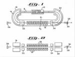
The extracted high-energy electron-beam can be directed into auxiliary components,
such as,
bending magnets and insertion devices (undulators or wigglers),
in storage rings and free electron LASERs,
supplying the strong magnetic fields,
perpendicular to the beam,
needed to convert the electrons' cinetic energy,
into quasi-monochromatic photons,
ranging from Infrared up to X-rays,
forming a tunable Free Electron LASER (FEL) [1].
First observed in synchrotrons,
synchrotron-light is now attempted to be produced by less expensive smaller specialized Compact Light Sources (CLS).
Compact light sources are not a substitute for large synchrotron and FEL light sources that typically also incorporate extensive user support facilities.
Rather they offer attractive,
complementary capabilities at a small fraction of the cost and size of large national user facilities.
In the far term they may offer the potential for a new paradigm of future national user facility.
The major R&D topics that could enhance the performance potential of both compact and large-scale sources include:
- The development of infrared (IR) LASER systems delivering kW-class average power with femtosecond pulses at kHz repetition rates.
These have application to ICS sources,
plasma sources,
and HHG sources.
- The development of LASER storage cavities for storage of 10-mJ pse and fsec pulses focused to micron beam sizes.
- The development of high-brightness,
high-repetition-rate electron sources.
- The development of continuous wave (cw) superconducting rf linacs operating at 4 K,
whilenot essential,
would reduce capital and operating cost.
- The development of Inverse Compton effect based Sources.
It is the purpose of this project to follow the innovation trail of quasi-monochromatic X-Ray sources,
from 1960 to 2015,
as it is reflected on relevant Industrial Property (IP) documents, in order to outline the miniaturization process that seems to lead towards tunable,
desk-top sized FELs,
into a modern Medical Imaging Laboratory.


