1.
Patients (Fig.
3)
◦ Retrospective review of medical records and cardiac CT scan
◦ Inclusion
- Patients who underwent MV annuloplasty from Jan 1995 to Dec 2013
AND
- Patients who underwent cardiac CT from Mar 2008 to Aug 2014
◦ Exclusion
-Patient with interval less than 1 year between CT and MV annuloplasty (n=13)
- Patients with rheumatic heart disease (n=6)
- Patients with no ring or other ring types (n=3)
- Patients with non-assessable CT image quality (n=2)
◦ A total of 45 patients were included in the final analysis
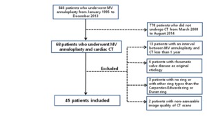
Fig. 3: A flow diagram of the study population.
2.
CT Image analysis
◦ Multiplanar reformatted images in a short-axis image of the MV annulus,
a long-axis view of the left ventricle,
and a 4-chamber view
◦Pannus formation,
valve leaflet thickening,
valve opening area,
diastolic opening angle,
and systolic coaptation angles of anterior and posterior mitral leaflets,
tenting height and mitral annular size (Fig.
4)
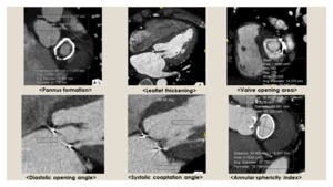
Fig. 4: Parameters for CT image analysis.
◦ Pannus formation around the annuloplasty ring
- Using short axis view of the mitral annulus
- Severity of pannus: both qualitative and quantitative methods (Fig.
5)
◦ Leaflet thickeness
- Both qualitative and quantitative methods (Fig.
5)
- Leaflet thickening: the maximal thickness of the MV leaflet >2mm
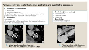
Fig. 5: Details for the assessment of pannus severity and leaflet thickening on CT.
◦ The opening area of the MV
- In cases in which multiphase data were available
- Measured at the maximal opening point of the valve tip
◦ The diastolic opening angle and systolic coaptation angles of the anterior and posterior mitral leaflets and the tenting height on systole
- 4-chamber view
◦ Assessment of mitral ring size
- The maximal and minimal diameters inside the annuloplasty ring
- The annular area
- The annular sphericity index: The maximal annular diameter/ minimal diameter
3. Data analysis
◦ Clinical data
- Etiology of original valve disease,
type and size of annuloplasty ring,
concurrent other valve operation or coronary artery bypass graft surgery,
and follow-up data for reoperation
◦ Transthoracic echocardiography (TTE) data
- Mean diastolic PG (MDPG),
MV area (MVA,
by pressure half time),
presence of mitral regurgitation,
and left ventricular ejection fraction
4. Statistical analysis
◦ Correlation between TTE parameter (PG,
PHT)and CT appearance (pannus,
leaflet thickening)
- Pearson correlation coefficient
◦ Comparison of CT findings between patients with normal PG and patients with elevated PG (mean-diastolic PG≥ 5mmHg)
◦ Comparison of incidence of functional MS on TTE,
presence of pannus,
and leaflet thickening on CT between the two ring types (Duran ring vs CE ring)
- Chi-square statistics or the Fisher exact test


