Purpose
To review from an educational point of view the radiologic manifestations of sinus venosus atrial septal defect.
Methods and Materials
Fivecases of sinus venosus atrial septal defect were collected from the report records of the Department of Radiology at Mayo Clinic,
Jacksonville,
Florida.
In collaboration with the Radiology Department of Clínica Universidad de Navarra,
we review the clinical behaviour,
imaging findings and final diagnosis of this uncommon disease.
Results
Prevalence
Atrial Septal Defect (ASD) isthe most common congenital heart defect to present in adulthood (1).
ASD accounts for 5% to 10% of all cases of congenital heart disease (2).
ASD can be caused by a defect of ostium secundum (80%),
ostium primum (10%),
unroofed coronary sinus (<1%) and sinus venosus (SVASD),
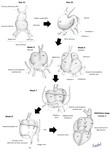
which is reportedto represent approximately 2-10% of all cases of ASD (2) (Figure 2).
Embryology
ASD can be classified in two types:
A) Direct communicatiom between the right and left atria: ostium primum...
Conclusion
Atrial septal defects are the most common congenital heart defect to present in adulthood and sinus venosus subtype represents 2-10% of all of ASDs.
Sinus venosus ASD (SVASD) is associated with partial anomalous pulmonary venos return.
A left-to-right shunt can be present,
which may lead to right ventricular volume overload,
development of elevated right ventricular pressures and possible subsequent development ofirreversiblepulmonary hypertension.
Surgical repair of of SVASD is usually indicated,
withsurvival rates similar to those expected for age and gendermatched controlled populations.
CTA and CMR...
References
1.
Carlos a.
Rojas.
Embryology and Developmental Defects of the interatrial Septum.
AJR Am J Roentgenol.
2010 Nov;195(5):1100-4.
2.
Hugh D.
White.
Imaging of adult atrial septal defects with angiography.JACC Cardiovasc Imaging.
2013 Dec;6(12):1342-5.
3.
Edward T.D.
Hoey.Multidetector CT assessment of partial anomalous pulmonary venous return in association with sinus venosus type atrial septal defect.
Quant Imaging Med Surg.
2014 Oct; 4(5): 433–434.
4.
JM Oliver.Sinus venosus syndrome: atrial septal defect or anomalous venous connection? A multiplane transoesophageal approach.Heart.
2002 Dec; 88(6): 634–638.
5.
Kafka...


