Purpose
Obstructive sleep apnea syndrome (OSAS) is the most common form of the breathing violation during sleep.
The syndrome often remains undiagnosed.
This condition is characterized by the presence of snoring,
repetitive partial or complete cessation of breathing during sleep,
long enough to reduce the level of oxygen in the blood,
rough sleep fragmentation and excessive daytime sleepiness.
The prevalence of OSAS is 5-7% of the population older than 30 years.
Severe forms of the disease affect approximately 1-2% of the specified group of persons.
Currently...
Methods and Materials
40 male patients with obesity (body-mass index was more than 30 kg/m2) and complaints of snoring were enrolled in the study.
All patients underwent polysomnography,
cardiorespiratory monitoring examinations and MRI (Phillips Achieva 3.0 T scanner with 16-channel head coil was used).
Patients had apnea-hypopnea index (AHI) of 52±17.
20 patients with minimal SpO2<75% formed the first group.
20 patients with minimal SpO2>75% were included in the second group.
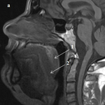
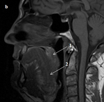
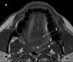
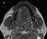
Area of maximum airway constriction (SmaxCA) at the level of retropalatal region,
volume of the soft palate...
Results
Our study showed area of maximum airway constriction (SmaxCA) at the level of retropalatal region of 0.5±0.2 cm2 and 0.7±0.2 cm2 for the first and the second groups,
respectively,
p<0.05.
Volume of the lateral walls was larger in patients with SpO2<75% (12.1±1.8 cm3 vs.
11.2±3.2 cm3) comparing with the second group,
p<0.05.
volume of the soft palate was comparable in two groups (9.1±1.5 cm3 vs.
7.9±2.5 cm3),
p=0.1.
In our study MR images of patients with different levels of oxygen saturation during sleep were examined....
Conclusion
Patients with obesity,
severe sleep apnea syndrome and oxygen blood saturation level less than 75% showed significantly smaller the upper airway section area and larger the volume of the lateral walls at the retropalatal region.
References
1. Chi L,
Comyn FL,
Mitra N,
et al.
Identification of craniofacial risk factors for obstructive sleep apnoea using three-dimensional MRI.
Eur Respir J 2011; 38:348–58.
2. Sutherland K.,
Richard J.
Schwab et al.
Facial Phenotyping by Quantitative Photography Reflects Craniofacial Morphology Measured on Magnetic Resonance Imaging in Icelandic Sleep Apnea Patients.
Sleep 2014; 37(5):959-968.
3. Amra B,
Peimanfar A,
Abdi E et al.
Relationship between craniofacial photographic analysis and severity of obstructive sleep apnea/hypopnea syndrome in Iranian patients.
J Res Med Sci.
2015; 20(1):...


