Purpose
Chest x-ray is widely used as screening before cardiac surgery.
The purpose ofthis study was to investigate how often thisleads to relevant information affecting clinical management.
Methods and Materials
1,136 patients from a tertiary referral center who were scheduled for cardiac surgery were included.
Surgeries were performed between 2011-2015.
To determine if the chest x-ray showed abnormalities and if these findings had clinical consequences,
radiology reports,
medical records and discharge letters were analyzed.
We also determined if any other chest x-rays or cardiac/chest computed tomography (CT) acquisitions were performed in the year prior to surgery and if there was a relation between an abnormal chest x-ray and postoperative complications.
Results
Baseline characteristics are provided in Table 1.
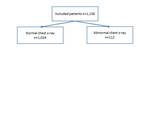
The chest x-ray was considered abnormal in 9.9% (112/1,136) patients (Figure 1).
The three most common abnormalities werea possible mass (42/1,136; 3.7%),
consolidation (19/1,136; 1.7%) or pleural effusion (42/1,136; 3.7%).
Additional test or referral to a specialist based on those findings occured in 17 patients (17/1,136; 1.5%).
The surgical strategy was altered in one patient,
due to extensive aortic calcifications noted on the chest x-ray and subsequent CT.
Surgery was postponed in two patients due to suspected previously...
Conclusion
The yield of a routine chest x-ray before cardiac surgery is very limited with regard to abnormal findings requiring a change in clinical management.
However,
an abnormal chest X-ray is associated with a prolonged stay in the intensive care unit.


