Population:
Between November 2015 and May 2016 there were 36 consecutive patients referred for evaluation of the peripheral arteries.
Contrast media protocol:
An uniform injection CM protocol was used.
A test bolus of 15 ml CM and 30 ml CM main bolus (Iopromide 300mgI/ml,
Ultravist,
Bayer; total iodine load was 13,5gI),
both followed by a 40 ml saline chaser were injected at 5ml/s injection rate with IDR 1,5gI/l.
Scan protocol:
Peripheral arteries were scanned on 3rd generation Dual Source CT scanner (Somatom FORCE,
Siemens Healthcare,
Forchheim,
Germany).
Scan protocol was individually tailored to each patient based on CM bolus transit time at the abdominal aorta and popliteal artery,
which were used to calculate scan time and scan delay from diaphragm to the forefoot.
Other scan parameters were kept constant with tube voltage of 70kV,
tube current of 90mAseff,
pitch 0.6,
collimation 0.6 and slice thickness 2/1.4mm.
Images were reconstructed with kernel Bf40.
Image Quality:
Image quality was assessed at the level of infrarenal aorta and then at five levels of peripheral arteries in both limbs,
left and right,
as follows: Common Iliac Artery (CIA),
Superficial Femoral Artery (SFA),
Popliteal Artery (PA),
Anterior Tibial Artery (ATA),
Dorsalis Pedis Artery (DPA).
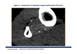
Objective image quality was evaluated as quantitative measurement of vascular attenuation in HU with manually placed circular regions of interest (ROI) and contrast to noise ratio (CNR) (see figure 1.).
Subjective image quality was evaluated with 4-point Likert scale (1=non-diagnostic,
2=diagnostic,
3=good,
4=excellent) independently by two experienced readers.


