Purpose
To compare radiation dose and image quality of turbo high-pitch dual-source computed tomography (DSCT),
dual-energy CT (DECT) and conventional single-source spiral CT (SSCT) for pulmonary CT angiography (CTA) on a 3rd generation dual-source CT,
respectively a 64-slice CT system.
Methods and Materials
Prospectively atotal of 120 patients matched for sex,
age,
and body-mass-index were identified for this study and divided in three groups of 40 patients each.
CTDIvol,
DLP,
subjective image quality as assessed by two readers (five-point scale: 1 = excellent; 2 = good; 3 = moderate; 4 = poor; 5 = non-diagnostic),
measured CT attenuation (HU) in three central and peripheral levels,
background noise (BN) and calculated signal-to-noise-ratio (SNR) were compared.
CT protocol settings of each patient group were chosen according to the manufacturer’s recommendations:...
Results
Mean CTDIvol and DLP were significantly lower (CTDIvol: 1 vs.
3: p<0.001; 2 vs.
3: p=0.002 / DLP: 1 vs.
3: p<0.001; 2 vs.
3: p<0.001) in group 3 (4.49±1.48 mGy / 161±55 mGycm) compared to group 1 (7.45±2.72 mGy / 252±96 mGycm) and group 2 (6.46±3.72 mGy / 228±136 mGycm).
Subjective image quality was rated good to excellent in >91% (110/120) with an interreader agreement of 90.3%.
The three protocols did not significantly differ in subjective image quality.
While group 3 presented with higher...
Conclusion
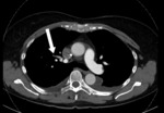
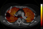
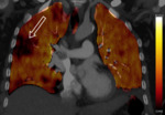
The use of third generation DECT in 90/Sn150 kV configuration allows for significant dose reduction in pulmonary CTA while providing excellent image quality and potential additional information by means of iodine perfusion maps (Figures 1 - 3).
References
Remy-Jardin M,
Pistolesi M,
Goodman LR,
et al.
Management of suspected acutepulmonary embolism in the era of CT angiography: a statement from the Fleischner Society.
Radiology.
2007;245:315–329.
Johnson TRC,
Krauss B,
Sedlmair M,
et al.
Material differentiation by dual energy CT: initial experience.
Eur Radiol.
2007;17:1510–1517.
Thieme SF,
Johnson TRC,
Lee C,
et al.
Dual-energy CT for the assessment of contrast material distribution in the pulmonary parenchyma.
Am J Roentgenol.
2009;193:144–149.
Bauer RW,
Kerl JM,
Weber E,
et al.
Lung perfusion analysis with dual energy...


