Purpose
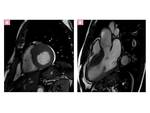
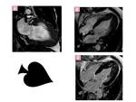
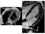
Resume in a practical guide for radiological use, how to perform a Cardiac Magnetic Resonance Imaging (CMRI) study in patients with suspected or confirmed Hypertrophic Cardiomyopathy (HCM), identifying the defining morphologic, physiologic and functional features of HCM and demonstrate the typical phenotypes of the disease.
Describe the interaction between different phenotypic patterns, functional and hemodynamic consequences, and potential adverse events and list the secondary signs of HCM changes during progression to the burned-out phase and medically refractory heart failure
Reveal the different patterns of late...
Methods and Materials
Hypertrophic cardiomyopathy (HCM)is the most common inherited cardiomyopathy, affecting one of every 500 adults. Representsthe most common cause of Sudden cardiac death (SCD)in young athletes due to malignant ventricular arrhythmias.HCM is caused by mutations in sarcomeric genes transmitted following an autosomal dominant pattern.There is agreat variability ofphenotypes.
The pathologic hallmarks of HCM aremyofibrillary disarray, interstitial fibrosis and abnormal dysplasia of intramural coronary arterioles (microvascular dysfunction).
The diagnosis requires exclusion of disease entities that may lead to inappropriate myocardial wall thickening of other etiologies (phenocopies).
Because...
Results
1-CMRISTUDY PROTOCOL:
CMRI tecniques let us a new perspective about the frequency, management, and prognosis of HCM.
-CINE SSFP MR IMAGING SEQUENCES:
(Cine Steady-state free precession) are used to evaluate:
-Cardiac function measurement
Biventricular volume quantification and function
Elevation of left ventricle ejection fraction(LVEF)
Sistolic disfunction often develops with end-stage HCM.
Assessment of global and segmental systolic thickening
-Morphological aspects:
Quantify the Myocardial thickness and mass accurately which are related to the diagnosis and prognosis of HCM:
Diagnostic criterion for HCM is a maximal LV...
Conclusion
-MRI has proven to be an important tool for the evaluation of patients suspected of having HCM.Cardiac MR imaging is useful for making a diagnosis and identifying the phenotypes of HCM , can also contribute to risk stratification because of its accurate measurement of LVH, detection of “high-risk” phenotypes, and identification of myocardial fibrosis
-An appropriate study protocol aimed at revealing the keys to the disease
and a deep understanding of the pathophysiology of the disease is essential to understand the role ofMRI paper in...
References
Rickers C, Wilke NM, Jerosch-Herold M, et al. Utility of cardiac magnetic resonance imaging in thediagnosis of hypertrophic cardiomyopathy. Circulation 2005;112(6):855–861.
Prasad K, Atherton J, Smith GC, McKenna WJ, Frenneaux MP, Nihoyannopoulos P. Echocardiographic pitfalls in the diagnosis of hypertrophic cardiomyopathy. Heart 1999;82(suppl 3):III8–III15.
Pennell DJ, Sechtem UP, Higgins CB, et al. Clinicalindications for cardiovascular magnetic resonance(CMR): Consensus Panel report. Eur Heart J 2004;25(21):1940–1965
Olivotto I, Cecchi F, Poggesi C, Yacoub MH et al. Patterns of disease progression in hypertrophic cardiomyopathy: an individualized approach to...


