Purpose
Coronary artery disease (CAD) is the single biggest cause of cardiac morbidity and mortality, while calcific aortic valve stenosis (AVS) is the third-leading cause of cardiovascular disease, placing a major burden onhealthcare systems throughout the world[1,2]. CAD develops as a chronic process of atherosclerotic plaque formation with progressive luminal narrowing. The natural history of these plaques varies, but most will form and evolve over years to decades, with atheroma remodelling and calcification, before any symptoms develop. Similarly,AVS often develops and progresses silently and by the...
Methods and Materials
This single-centre retrospective study included 400 consecutiveunenhanced non-ECG-gated chest CT examinations of patients between the ages of 40 and 69 years over a period of three months. Exclusion criteria included previous cardiac intervention (coronary stent, coronary artery bypass graft or valve replacement) and repeat scans inthis time period. All scans were performed using volumetric acquisitionswith slice thickness of 1mmon a multislice CT scanner. The kV median and interquartile range (IQR) was 120 [120-130] and mA adjusted according to patient factors. The mediastinum window [WW: 400,...
Results
Out of the 400 consecutive CT chest examinations, 375 were eligible for inclusion in the study.169 (45%) were positive for coronary artery and/or aortic valve calcification (Table 1), while 206 (55%) were negative for both.
Coronary artery calcification
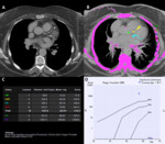
The CT chest scans of 162 (43%) patients were positive for CAC, out of which 36 (10%) were also positive for aortic valve calcification. 71 (44%) patients were female and 91 (56%) were male. The total median and interquartile range (IQR) age was 63 [57-67] years, which...
Conclusion
This study shows that coronary artery calcification (CAC) is a frequent incidental finding in middle-aged adults undergoing chest CT, and over a quarter of patients with CAC showed moderate/severe disease. In only 35% ofthe cases was the presence of CAC mentioned by the reporting radiologist, which is slightly lower than in other studies[4]. However, a relatively low number of unreported CAC was categorised as moderate or severe disease.The traditional management of moderate and severe coronary artery calcification based on the Agatston categories includes aggressive risk...
References
1.Benjamin EJ,Virani SS,Callaway CW, et al. Heart Disease and Stroke Statistics — 2018 update: a report from the American Heart Association. Circulation. 2018;137(12):e67-e492.
2.Pibarot P, Dumesnil JG. New concepts in valvular hemodynamics: implications for diagnosis and treatment of aortic stenosis. TheCanadian Journal of Cardiology.2007;23 Suppl B(Suppl B):40B–47B.
3.Pawade T, Clavel MA, Tribouilloy C, et al. Computed tomography aortic valve calcium scoring in patients with aortic stenosis.Circulation: Cardiovascular Imaging. 2018;11(3):e007146.
4.Hecht HS, Cronin P, Blaha MJ,et al.2016 SCCT/STR guidelines for coronary artery calcium scoring of noncontrast...


