Out of the 400 consecutive CT chest examinations, 375 were eligible for inclusion in the study. 169 (45%) were positive for coronary artery and/or aortic valve calcification (Table 1), while 206 (55%) were negative for both.
Coronary artery calcification
The CT chest scans of 162 (43%) patients were positive for CAC, out of which 36 (10%) were also positive for aortic valve calcification. 71 (44%) patients were female and 91 (56%) were male. The total median and interquartile range (IQR) age was 63 [57-67] years, which was similar in both genders with female 62 [57-66] years and male 63 [56-67] years. The median visual score was 2 [1-4] and median Agatston score was 95 [20-322].
The degree of calcium severity, as assessed by Ordinal visual score, was found to be 117 (72%) mild, 28 (17%) moderate and 17 (11%) severe calcification, with more than a quarter showing moderate/severe calcification (Figure 1). In only 57 (35%) of the reports was presence of coronary calcium mentioned by the reporting radiologist. Of those not reported, 87 (83%), 15 (14%) and 3 (3%) were in the mild, moderate and severe category, respectively.
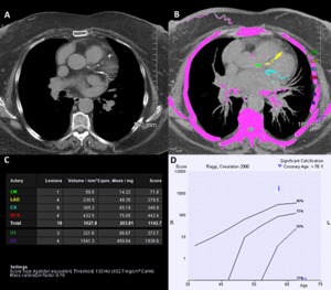
A high degree of association was observed between the severity ranking of CAC by visual and Agatston score, with 135 (83%) of severity rankings in the same category. The accuracy was 90%, 85%, and 91% in the mild, moderate, and severe CAC severity categories, respectively. This showed strong correlation (r=0.82, p<0.0001) between the visual and Agatston scores (Figure 2) using Spearman rank correlation coefficient. Figure 3 shows an example of severe coronary artery calcification both by visual and Agatston score.

Fig. 3: Unenhanced CT chest of a 62 year old male displaying calcification in the left main stem (LMS), left anterior descending (LAD), and circumflex (LCX) arteries, with severe calcification by Ordinal (9) and Agatston (1143) scores. Note severe calcification of a large OM branch, which was not included in the Ordinal score and represents a limitation of the method. a. Original CT chest used to calculate the Ordinal score. b. The same CT chest study with syngo.via* software for the calculation of Agatston score. c. Agatston score table automatically generated by the software* with values per coronary artery territory. Also included are the scores for aortic valve (U1) and mitral annnulus (U2) calcification. d. Graphic representation of distribution of Agatston scores in age and gender matched patients. Note the Agatston score places this patient above the 90th percentile for matched age and gender.
Aortic valve calcification
43 (12%) of the scanned adults had aortic valve (AV) calcification with 36 (84%) having coronary artery calcification concomitantly. 23 (53%) of the patients were female and 20 (47%) were male with the median age of 64 [61-67] years.
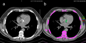
The median Agatston score was 49 [25-206], with the majority of patients (29, 67%) with Agatston score of less than 150. 14 (33%) patients had Agatston score of more than 150 with 4 (9%) of them recording a value above 500. Figure 4 shows an example of severe aortic valve calcification.

Fig. 4: a. Unenhanced CT chest of a 62 year old male with severe calcification by visual scoring. b. The same study with syngo.via software for calculation of the Agatston score, which was 1404 (severe).
In 37 (86%) of the cases, the visual and Agatston scores were in agreement. Only in 5 cases was the presence of aortic valve calcification mentioned in the report, with 4 of those patients having Agatston score above 150. 8 patients with Agatston score above 150 had no mention of aortic valve calcification in their report.


