Purpose
The purpose of our study is to evaluate the prevalence of structural lesions on 3.0T MR imaging of patients presenting with nystagmus secondary to MS,
to examine the clinicopathological correlations between nystagmus and lesions in the central nervous system (CNS),
and to evaluate whether nystagmus could occur as a result of cortical lesions or disruption to the tracts connecting the cortical centres to the brainstem.
Methods and Materials
This observational cross-sectional study was conducted with the approval of the Ethics Committee of St.
James’s Hospital.
All patients gave written informed consent to the study.
We selected 30 patients with nystagmus secondary to MS,
who attended the neurology outpatient service.
All patients with an established diagnosis of MS who had nystagmus on clinical examination were considered to be eligible.
Exclusion criteria were as follows: any patient who was not voluntarily willing to undergo the scan or had other objections,
pregnant patients,
patients under the...
Results
Out of thirty patients,
twenty two were women (73%) and eight men (27%).
Ages ranged from 20 years to 60 years.
The types of nystagmus were as follows: horizontal bidirectional gaze-evoked nystagmus (50%); unidirectional gaze evoked nystagmus (26%); pendular nystagmus (3%),
torsional up gaze nystagmus (3%); and combined nystagmus (18%).
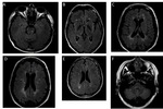
Representative images are shown in Figure 2.
Anatomical Location of Lesions
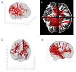
FA values were calculated for the twenty two patients who underwent tractographic analysis starting from various cortical eye fields leading up to the brainstem....
Conclusion
According to conventional description,
nystagmus originates from lesions in the neural integrator circuit involving the brainstem,
vestibular nuclei and cerebellum.
Studies have shown that infratentorial lesions are expected in up to 40% of patients with MS (9-11).
We expected to find higher numbers in our study group,
because we used 3T MRI which is more sensitive in identifying lesions.
If the traditional theory is true,
then we should have observed infra tentorial and brainstem lesions among all patients in the study group.
Instead we found...
References
Conturo,
T.E.,
N.F.
Lori,
T.S.
Cull,
E.
Akbudak,
A.Z.
Snyder,
J.S.
Shimony,
R.C.
McKinstry,
H.
Burton,
and M.E.
Raichle.
Tracking neuronal fiber pathways in the living human brain.
Proceedings of the National Academy of Sciences of the United States of America,
1999.
96(18): p.
10422.
Minneboo A,
Barkhof F,
Polman CH,
Uitdehaag BM,
Knol DL,
Castelijns JA.
Infratentorial lesions predict long-term disability in patients with initial findings suggestive of multiple sclerosis.
Arch Neurol 2004;61:217-221.
Sailer M,
O'Riordan JI,
Thompson AJ,
et al.
Quantitative MRI in...


