ECR 2011 / C-1911
Visualization of visual pathway disruption in patients with nystagmus associated with multiple sclerosis: value of DTI at 3T
Congress:
ECR 2011
Poster Number:
C-1911
Type:
Scientific Paper
Keywords:
Neuroradiology brain, MR-Diffusion/Perfusion, MR, Physiological studies, Inflammation
Authors:
R. Dunne, P. Iyer, J. F. Meaney, A. Fagan, K. Curran, N. Colgan, K. Curran, G. Boyle, J. Redmond; Dublin/IE
DOI:
10.1594/ecr2011/C-1911

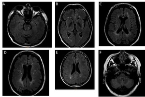
Fig. 1:
Anatomical Location of Whitte Matter Lesions
 Brainstem; (B) temporal atrophy; (C) frontal & periventricular; (D) temporal; (E) occipital; (F) cerebellar")
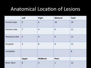
Fig. 2:
3T MRI Images of patients in the study showing lesions in various areas:
(A)...
 Disruption in frontal area; (B) left-sided fiber disruption (C) bi frontal tract disruption (D) elevation view with bilateral frontal disruption.")
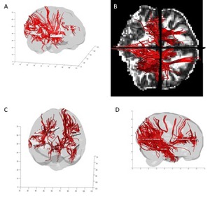
Fig. 3:
Examples of fiber tract disruption represented in different views observed on...