Purpose
This study was performed to provide preliminary clinical feasibility of a noninvasive hybrid imaging modality,
Imagio,
that uses combination of real-time laser opto-acoustic and ultrasound for improving diagnostic accuracy in the evaluation of breast masses. The system characterizes and differentiates breast tumors based on the concentration of blood and its oxygen saturation in the tumor angiogenesis while also showing morphological information based on traditional ultrasound imaging methods. Opto-acoustic imaging uses pulses of laser light in the near-infrared spectral range to illuminate tissues and detects the...
Methods and Materials
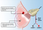
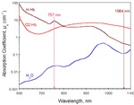
Laser illumination at the wavelength of 757 nm provides contrast based mainly on the hypoxic blood of breast carcinomas,
while a wavelength of 1064 nm produces contrast dominated by the enhanced water content and normally oxygenated blood in benign fibroadenomas.
Detection of the resulting ultrasound signals with a commercial handheld ultrasound probe preserves quantitative information about the tumor optical absorption. Two opto-acoustic measurements yield solutions for the concentrations of hemoglobin and oxy-hemoglobin in pixels within the field of view. In the same location,
ultrasound images...
Results
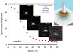
Experiments in an optically scattering phantom mimicking a blood vessel in the breast demonstrated capability of theopto-acoustic imaging system to detect blood vessels at the depth of at least 6 cm.After the system was calibrated in phantoms,
a feasibility study was performed on female patients with breast masses having BIRADS scores of 4 and 5 and who were scheduled for biopsy.
Initial studies on 32 patients demonstrated that the combined opto-acoustic / ultrasound imaging system can detect areas of high optical absorption in the region...
Conclusion
The combination of optically-induced functional contrast and acoustically generated high resolution anatomical imaging in a novel breast cancer imaging modality demonstrated clinical feasibility and the potential for noninvasive diagnostics.
Opto-acoustic system demonstrated depth of imaging comparable with that of ultrasound.
Coregistered ultrasound and opto-acoustic images provide complimentary morphological and functional images. This new dual modality is envisioned as an adjunct to X-ray mammography that provides functional and anatomical maps.
References
S.A.
Ermilov,
T.
Khamapirad,
A.
Conjusteau,
R.
Lacewell,
K.
Mehta,
T.
Miller,
M.H.
Leonard,
A.A.
Oraevsky: Laser Optoacoustic Imaging System for Detection of Breast Cancer,
J Biomed Opt.
2009; 14(2): 024007 (1-14).
S.
Ermilov,
M.
Fronheiser,
H.-P.
Brecht,
R.
Su,
A.
Conjusteau,
K.
Mehta,
P.
Otto,
A.
Oraevsky: Real-time optoacoustic imaging of breast cancer using an interleaved two laser imaging system coregistered with ultrasound.
Proc.
SPIE 2010; 7564: 75641W.
Personal Information
Dr.
Pamela Otto is a Professor of Radiology at the University of Texas Health Science center in San Antonio.


