ECR 2013 / C-0595
A new method for radiation dose reduction at cardiac CT with multi-phase data-averaging and non-rigid image registration: preliminary clinical trial
Congress:
ECR 2013
Poster Number:
C-0595
Type:
Scientific Exhibit
Keywords:
Cardiac, Cardiovascular system, CT, CT-Angiography, Image manipulation / Reconstruction, Comparative studies, Technical aspects, Cardiac Assist Devices
Authors:
F. Tatsugami, T. Higaki, M. Kiguchi, S. Date, K. Awai; Hiroshima/JP
DOI:
10.1594/ecr2013/C-0595

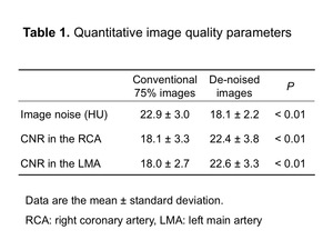
Table 1
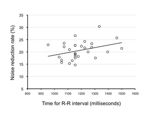
 increased in proportion to the time for the R-R interval (msec) (Pearson correlation, r = 0.42, P = 0.02). Since the noise reduction rate increased in proportion to the time for the R-R interval, we suggest that our method is applicable in patients with a lower heart rate.")
Fig. 3:
Noise reduction rate (%) increased in proportion to the time for the R-R...
.")
Fig. 4:
Image quality score for the right coronary artery (RCA).
.")
Fig. 5:
Image quality score for the left anterior descending coronary artery (LAD).
.")
Fig. 6:
Image quality score for the left circumflex coronary artery (LCX).
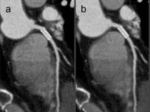
 Conventional 75% image. The image noise was 22.3 HU and CNR in the left main coronary artery was 19.4 and the image quality was rated as good. (b) De-noised image. The image noise was 17.4 HU and CNR in the left main coronary artery was 24.5 and the image quality was rated as excellent.")
Fig. 7:
A 51-year-old man with a body mass index of 22.0 kg/m2 who was referred for...
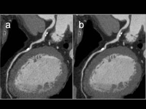
 Conventional 75% image. The image noise was 25.2 HU and CNR in the left main coronary artery was 16.3 and the image quality was rated as good. (b) De-noised image. The image noise was 20.5 HU and the CNR in the left main coronary artery was 20.1 and the image quality was rated as excellent.")
Fig. 8:
A 76-year-old man with atypical chest pain and risk factors for coronary artery...