Keywords:
Metastases, Metabolic disorders, Cancer, Imaging sequences, Diagnostic procedure, Computer Applications-Detection, diagnosis, Ultrasound, MR, CT, Paediatric, Neuroradiology brain, Abdomen
Authors:
Y. Tachibana1, R. Kishimoto1, T. Omatsu1, S. Kandatsu2, A. Hasegawa1, M. Koto1, R. Takagi1, T. Obata1, H. Tsuji1; 1Chiba/JP, 2Chiba-Ken/JP
DOI:
10.1594/ecr2013/C-0959
Methods and Materials
Patients:
Clinical databases were retrospectively searched for patients who underwent both SWV measurement and MRI within a 90-day interval to investigate their cervical lymph nodes with the suspicion of metastasis.
Patients with lymph nodes not matching clearly between the two modalities were excluded.
Finally,
6 patients with lymph nodes considered metastatic (pathologically and/or clinically: metastasis group) and 17 patients without obvious metastatic nodes (pathologically,
or at least 6 months of clinical observation: control group) met the criteria.
The largest node from each patient was selected as object.
SWV measurement:
Acuson S2000 (SIEMENS AG,
Germany) with 9MHz linear probe was used.
All exams were done by the same trained radiologist.
The region-of-interest (ROI) was carefully set on the center of the lymph nodes (Figure 2: B),
and SWV was measured 1-3 times for each lymph node (3 times for 20 lymph nodes,
twice for one lymph node,
and once for one lymph node).
When SWV was measured twice or more,
the median value was assessed in this study.
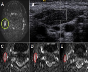
Fig. 2: A 71-year-old man with a pathologically proven metastatic lymph node (adenocystic carcinoma). A; Diffusion-weighted image shows multiple enlarged lymph nodes bilaterally. The largest lymph node was selected as object (green circle). B; Shear wave velocity (SWV) measurement in the corresponding lymph node. The rectangle indicates the region of interest (ROI). C-E; The ROIs were set manually to evaluate apparent diffusion coefficient parameters; up to three slices were selected for the ROI when the lymph node was visible in multiple slices (as in this sample case). These ROIs were grouped and were considered as a single ROI.
ADC measurement:
Achiva 1.5T (Philips Medical Systems,
Netherlands) with an 8-channnel head coil was used.
ADC was measured from DWI underwent as a part of routine MRI study.
The main parameters were as follows: TR 4000msec,
TE 52msec,
average 2,
matrix 144 x 93,
FOV 230mm,
thickness 5mm,
bandwidth 1425Hz/pix,
b-value 0, 1000sec/mm2 ,scantime 1min12sec.
ROI settings and comparison:
The average,
median,
and standard deviation (SD) of ADC were acquired in a ROI-based study (Figure 2: A,
C-E).
The parameters of ADC and SWV were compared between the control and metastasis groups (unpaired t-test).
The coefficients of correlation between the parameters of ADC and SWV were also evaluated in each group separately,
and also in the group of all subjects. p<0.05 was considered significant for the comparisons.

. A; Diffusion-weighted image shows multiple enlarged lymph nodes bilaterally. The largest lymph node was selected as object (green circle). B; Shear wave velocity (SWV) measurement in the corresponding lymph node. The rectangle indicates the region of interest (ROI). C-E; The ROIs were set manually to evaluate apparent diffusion coefficient parameters; up to three slices were selected for the ROI when the lymph node was visible in multiple slices (as in this sample case). These ROIs were grouped and were considered as a single ROI.")