ECR 2013 / C-1007
Imaging characteristics of thoracic sarcomas – an illustration of interesting cases
Congress:
ECR 2013
Poster Number:
C-1007
Type:
Educational Exhibit
Keywords:
Neoplasia, Education and training, Cancer, Diagnostic procedure, PET, MR, CT, Thorax, Oncology, Lung
Authors:
D. Tzias, H. J. Cassidy, D. Douraghi-Zadeh, A. Devaraj, A. Nair; london/UK
DOI:
10.1594/ecr2013/C-1007
 Large left sided mass with osseous destruction of the posterior ribs and (b) posterior mediastinal mass with right sided rib scalloping, indicating the mass may have been present for some time. References: Royal Brompton Hospital")
Fig. 1:
Plain chest radiographs of primary chest wall sarcomas demonstrating; (a) Large...

Fig. 2:
Heavily calcified, irregular mass arising from the anterior chest wall with rib...

Fig. 3:
Axial CT of the chest demonstrating metastatic disease from a uterine...

Fig. 4:
Axial CT scan demonstrating a calcified metastasis in the left lower lobe. The...
 showing a treated osteosarcoma of the right chest wall post chemothearapy and radiotherapy; Coronal PET(b) of the same lesion showing low grade uptake of FDG, suggesting partial treatment response. By comparison, on lung windows(c) coronal CT shows a calcified osteosarcoma lung metastasis with avid FDG uptake on PET(d). References: Royal Brompton Hospital")
Fig. 5:
Coronal non contrast CT(a) showing a treated osteosarcoma of the right chest...
 demonstrates the cystic lesion in the right lower lobe. Plain radiograph(b) shows the cyst which has ruptured and contains a fluid level. References: Royal Brompton Hospital")
Fig. 6:
Patient, treated for left sided pneumothorax, with bilateral lower lobe...

Fig. 7:
Axial T2w MRI showing a right anterior chest wall mass that is seen to invade...
 demonstrates the separation of the left subclavian artery(characterised by signal void(arrowhead)), from the mass, better than CT(b). (d)Note the interscalene fat pad in the normal position on the right(arrow), but displaced on the left(arrow). If this fat pad is obliterated, then invasion of the brachial plexus should be suspected[4]. This is easier demonstrated on MRI than CT(c). References: Royal Brompton Hospital")
Fig. 8:
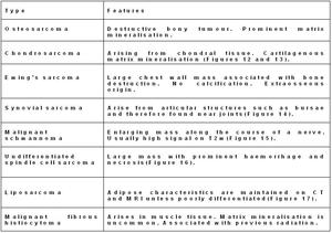
Osteosarcoma invading the left lung apex and pleura but sparing the subclavian...
 and T1w(b) of an undifferentiated spindle cell sarcoma. The mass is seen to invade the chest wall, necessitating reconstruction surgery, but the pectoralis muscle is spared. Note the fat plane between the mass and the muscle on CT and MRI(arrows). References: Royal Brompton Hospital")
Fig. 9:
Axial contrast enhanced CT(a) and T1w(b) of an undifferentiated spindle cell...
 and contrast enhanced CT(b) of a chondrosarcoma returning high T2w signal, arising from the right costal cartilage. Note the clearer depiction of the plane between the mass and the liver on MRI which demonstrates lack of liver invasion. References: Royal Brompton Hospital")
Fig. 10:
Axial T2w MRI(a) and contrast enhanced CT(b) of a chondrosarcoma returning high...
 and T2w(b) MRI demonstrates widening of the right neural foramen and clear invasion of the spinal canal. Note the scalloping that the mass is causing to the vertebral body. References: Royal Brompton Hospital")
Fig. 11:
Chondrosarcoma. Axial T1w(a) and T2w(b) MRI demonstrates widening of the...

Fig. 12:
Chondrosarcoma. Axial, contrast enhanced CT scan showing a heavily calcified...
 CT demonstrating typical “arc like” calcification of chondrosarcoma. References: Royal Brompton Hospital")
Fig. 13:
Chondrosarcoma with mediastinal and intraspinal extension. Axial, non...

Fig. 14:
Synovial sarcoma. Axial, contrast enhanced CT scan showing a chest wall mass...
 and low signal on T1w(a) MRI imaging due to myxoid tissue composition. Often demonstrates dots of low signal(arrows) on T2w imaging which represents collagen and condensed Schwann cells. References: Royal Brompton Hospital")
Fig. 15:
Malignant scahwanoma. Large mass arising from the rib usually high signal in...

Fig. 16:
Undifferentiated spindle cell sarcoma. Coronal, contrast enhanced CT scan...
, Axial post gadolinium T1w fat suppressed images showing a posterior chest wall mass with signal suppression and enhancing septations. (b) T2w axial imaging showing high signal return from the mass. (c) Axial, contrast enhanced CT scan showing the low attenuation fatty mass with septations. Biopsy showed well differentiated liposarcoma. The presence of thick septae, nodules and large mass size favours liposarcoma over lipoma. Note the intercostal chest wall invasion(arrow). References: Royal Brompton Hospital")
Fig. 17:
Liposarcoma. (a), Axial post gadolinium T1w fat suppressed images showing a...

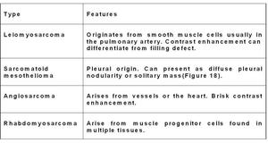
Fig. 18:
Axial post contrast, lung window, CT scan slice showing a soft tissue mass...