ECR 2013 / C-1479
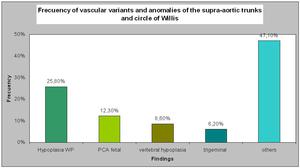
Variants and vascular anomalies of supra-aortic trunks and circle of Willis: A Pictorial Review.
Congress:
ECR 2013
Poster Number:
C-1479
Type:
Educational Exhibit
Keywords:
Computer Applications-Detection, diagnosis, CT-Angiography, Catheter arteriography, Neuroradiology brain, Anatomy, Congenital
Authors:
M. M. Padilla Deza1, D. Rodriguez2, L. Aja Rodriguez3, P. Mora Montoya3, Y. P. Velasco Díaz4, A. Muntané Sánchez4; 1Hospitalet de Llobregat, Ba/ES, 2El Vendrell/ES, 3Barcelona/ES, 4Hospitalet de Llobregat /ES
DOI:
10.1594/ecr2013/C-1479

Fig. 18

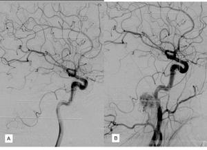
Fig. 19:
Fenestration of basilar artery asociated with aneurysm.
A. Before...

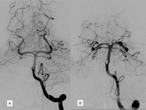
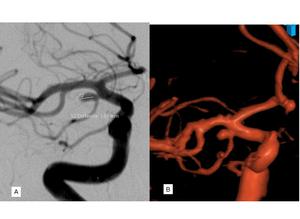
Fig. 20:
Azygos. DSA.
A.AP view. Right carotid artery angiogram.
B.AP view. Left...

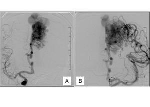
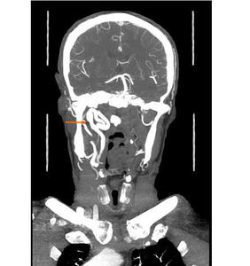
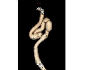
Fig. 21:
Retropharyngeal carotid associated with dissecant aneurysms

Fig. 22:
Retropharyngeal carotid associated with dissecant aneurysms

Fig. 23:
Temporal anterior branch originated in ICA, associated with aneurysm

Fig. 24:
A. PICA with origin in ICA.
B. In this patient was asociated with AVM.