ECR 2013 / C-1667
Pediatric Tuberculosis: pictorial review of radiologic findings
Congress:
ECR 2013
Poster Number:
C-1667
Type:
Educational Exhibit
Keywords:
Thorax, Lung, Lymph nodes, CT, Conventional radiography, Computer Applications-Detection, diagnosis, Cavitation, Infection
Authors:
M. Occhipinti1, A. R. Larici1, A. del Ciello1, P. Franchi1, E. Devicienti2, L. Calandriello1, F. Maggi1, L. Bonomo1; 1Rome/IT, 2Roma/IT
DOI:
10.1594/ecr2013/C-1667
 shows a large cavity (star) within consolidation in right upper lobe (RUL). Axial CT scan at lung window setting (b) confirms the cavitating process in RUL, showing various cavities in the same lobe and an inner air-fluid level (arrow). Sagittal MPR image allows a better characterization of the cavity, which is unique and presents thick internal septa.")
Fig. 2:
Infant disease. CXR anteroposterior view (a) shows a large cavity (star) within...
. CXR posteroanterior view (a) shows multiple cavitations (asterisks) over both upper lobes, with a significant decrease in volume of RUL, as demonstrated by minor fissure elevation (yellow arrows). Poorly defined consolidations associated with cavitations (asterisks) and bronchiectasis in the upper third of the left hemithorax are also present. Coronal MPR image at lung window setting (b) confirms consolidations (short arrow), cavitations (asterisks) and bronchiectasis (long arrows) over both upper lobes.")
Fig. 3:
Adult-type cavitating disease (1). CXR posteroanterior view (a) shows multiple...
. In the same boy of Fig. 3 axial CT scans at lung window setting (c-d) demonstrate cystic and cylindrical bronchiectasis (asterisks) surrounded by parenchymal consolidations over both upper lobes, more evident at right side. Bronchial wall thickening (yellow arrow), poor defined small solid nodules and little cavitations (green arrow) are also present. Coronal minIP reformation image (e) exalts bronchiectasis, helping radiologist to distinguish between true cavities and cystic bronchial dilation.")
Fig. 4:
Adult-type cavitating disease (2). In the same boy of Fig. 3 axial CT scans at...

Fig. 5:
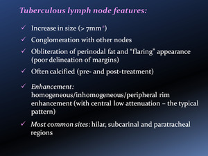
Different types of TB lymphadenopathies in children.
 shows a multilobulated consolidation of the lateral basal segment of right lower lobe (RLL). Axial CT image at mediastinal window setting (b) shows an hylar enlarged lymph node (yellow arrow). Oblique MPR image (c) better depicts the consolidations along the bronchovascular bundle (orange arrows), suggesting a bronchogenic spread. Bronchogenic spread of disease occurs when an area of caseous necrosis liquefies and communicates with the bronchial tree. Typically it involves lower lung zones, as in this case.")
Fig. 9:
Axial CT image at lung window setting (a) shows a multilobulated consolidation...
 shows right pleural effusion (arrow). Axial CT scan at lung window setting (b) shows only a linear parenchymal consolidation in the posterior basal segment of RLL, without a severe lung disease.")
Fig. 10:
Chest CT scan at mediastinal window setting (a) shows right pleural effusion...
 show consolidation of the apicodorsal segment of LUL (star). Axial enhanced CT scans at mediastinal window setting (b-c) show hylar enlarged lymph nodes (arrow in b) and the inhomogeneous low attenuation of the expansile pneumonia (c).")
Fig. 11:
Chest CT scans at lung window setting (a) show consolidation of the apicodorsal...
 shows mild opacifications in the right lung (stars). Axial CT scans at lung window setting localize the alveolar consolidations (asterisks) in different lobes: lateral and posterior basal segment of RUL with air-bronchogram (b), medial segment of middle lobe (ML) (c) and anterior basal segment of RLL (d). Axial enhanced CT scan at mediastinal window setting (e) shows mild calcifications within a consolidation and hylar lymph nodes calcifications (arrow).")
Fig. 12:
CXR (a) shows mild opacifications in the right lung (stars). Axial CT scans at...
 and at mediastinal window setting (b) show the typical apical cavitation (star), with thick wall (purple arrow) and an inner air-fluid level (green arrow), consistent with superinfection. Enlarged and inhomogeneous hylar and subcarinal nodes are also evident (yellow arrows) (c). Sagittal MPR (d) shows the cavitation within expasile consolidation in the apicodorsal segment of left upper lobe (LUL), with fissural buldging (orange arrow) and other parenchymal consolidations in the left lower lobe (LLL).")
Fig. 13:
Axial CT scans at lung window setting (a) and at mediastinal window setting (b)...
 1 week after CT scan [Fig. 13] shows reduction of consolidation and cavitation of LUL (arrow), even more reduced 1 month later.")
Fig. 14:
CXR anteroposterior view (e-f) 1 week after CT scan [Fig. 13] shows reduction...
 show compression of trachea (orange arrow) and carina (green arrow) associated to atelectasis of the apical and anterior segment of RUL (star). Axial enhanced CT scan at the same levels (b-d) show large, conglomerate enhancing nodes with central low attenuation areas, as for central necrosis (yellow arrows). (e-f) Other pathologic lymph nodes are also present in the supraclavicular and hylar regions (pink arrow).")
Fig. 15:
Axial CT scans at lung window setting (a-c) show compression of trachea (orange...
 shows complete opacification of left hemithorax, with mild shift of mediastinal structures to the right (yellow arrow).
Axial CT images at lung window setting (b-c) show: a wide cavity with internal septa in the left apex (b), lingular and LLL consolidation with air-bronchogram (orange arrow), loculated pneumothorax in the anterior part of the right hemithorax (star). Axial non-enhanced CT scan at mediastinal window setting (d) shows pleural effusion (green arrow) and consolidation of the adjacent parenchyma.")
Fig. 16:
CXR anteroposterior view (a) shows complete opacification of left hemithorax,...
 show thickening of visceral and parietal right pleural surfaces (arrows) separated by the presence of fluid (star), representing the ‘‘split pleura’’ sign. Axial CT image at lung window setting (c) shows no underlying parenchymal abnormalities.")
Fig. 17:
Axial enhanced CT images at mediastinal window setting (a-b) show thickening of...
 and bilateral pleural effusions (stars). Atelectasis of basal-posterior segment of both lower lobes (RLL>>LLL) is also present. References: Burrill J et al (2007) Tuberculosis: a radiologic review. RadioGraphics 27: 1255-1273")
Fig. 18:
Axial enhanced CT scan at mediastinal window setting demonstrates a thickened...
 shows multiple disseminated nodules in both lungs, calcified right hylar nodes (arrow) and the loss of sharpness of the right cardiac profile (star), due to partial consolidation of the ML. Chest CT scans at lung window setting (b-c) better depict the diffuse involvement of both lungs through multiple 2-3 mm nodules. A partial consolidation of the ML with inner calcifications is also present. Axial non-enhanced CT scans at mediastinal window setting (d-e) show calcified nodes in hylar, subcarinal and right lower paratracheal regions (arrows).")
Fig. 19:
CXR anteroposterior view (a) shows multiple disseminated nodules in both lungs,...
 shows a right latero-cervical opacity (arrows), due to laterocervical adenopathies, confirmed at CT scan (b). No signs of compression/infiltration of adjacent structures are present.")
Fig. 21:
CXR anteroposterior view (a) shows a right latero-cervical opacity (arrows),...
 show enlarged lymph nodes in the intercavo-aortic and left paraaortic regions (yellow arrows).
A calcified Ghon focus in the superior lingular segment (green arrow) associated with calcified hylar and subcarinal nodes is also present (orange arrows) (c-d) . These features are typical seen in post-primary tuberculosis.")
Fig. 22:
Axial CT images at abdominal window setting (a-b) show enlarged lymph nodes in...
 and anteriorly to the tip of temporal hornes of lateral ventricula (green arrows). High-intensity signals in the right basal ganglia (orange arrow) are also noted.")
Fig. 23:
T1-weighted MRI after contrast administration show florid leptomeningeal...
. There is an accompanying cold abscess overlying the right temporo-occipital region (curved arrow). References: Harisinghani MG et al. (2000) Tuberculosis from Head to Toe. RadioGraphics 20: 449-470")
Fig. 24:
Bilateral tuberculous mastoiditis. High-resolution CT scan of the temporal bone...
 shows consolidation of LLL (retrocardiac opacity) and hyperinflation of LUL. A mild diffuse opacity in the controlateral lung is also present. Axial CT scans at lung window setting (b-c-d), performed 2 days after CXR, confirm the difference of attenuation between the two lungs, the consolidation of LLL and the compression of the left main bronchus by enlarged nodes. Axial enhanced CT scan at mediastinal window setting (e) exalts lymphadenopathies in subcarinal and hylar regions (asterisks), partially obstructing lingular lobar bronchus. Occlusion of LLL bronchus by dense secretions may also be noted (arrow).")
Fig. 25:
CXR anteroposterior view (a) shows consolidation of LLL (retrocardiac opacity)...
 performed after 1 month of anti-TB therapy shows initial regression of LLL consolidation (arrows) and reduction of LUL hyperinflation. This finding indirectly indicates a reduction of size of lymph nodes.
Axial CT scan at lung window setting (b) shows residual little subpleural consolidations in the LLL (arrowhead), without differences in parenchymal attenuation between the two lungs. Axial CT images at mediastinal window setting (c-d-e) highlight the presence of soft calcifications (arrow) within consolidations and the regression of necrotic lymphadenopathies previously seen in left hylar and subcarinal regions.")
Fig. 26:
CXR anteroposterior view (a) performed after 1 month of anti-TB therapy shows...