ECR 2013 / C-1861
Combined approach to atrial and ventricular function for assessment of diastole through MRI: Hypertrophic Cardiomyopathy (HCM) vs Healthy Controls (HC).
Congress:
ECR 2013
Poster Number:
C-1861
Type:
Scientific Exhibit
Keywords:
Congenital, Imaging sequences, MR, Cardiac
Authors:
G. Gentile1, G. Aquaro2, E. Grassedonio1, G. Todiere2, P. Toia1, L. La Grutta1, C. Tudisca3, M. Lombardi2, M. Midiri1; 1Palermo/IT, 2Pisa/IT, 3Palermo, PA/IT
DOI:
10.1594/ecr2013/C-1861

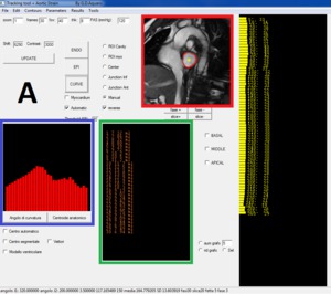
Fig. 1:
Screenshot of the software used for atrial function analysis

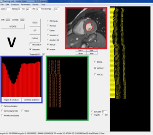
Fig. 2:
Screenshot of the software used for ventricular function analysis
 and derived dV/dT curves (C-D) References: Giovanni Gentile MD")
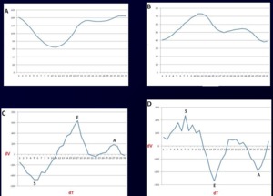
Fig. 3:
Ventricular and atrial Volume/Time curves (A-B) and derived dV/dT curves (C-D)