Keywords:
Artifacts, Aneurysms, Stents, Embolisation, Computer Applications-3D, CT-High Resolution, CT-Angiography, Cone beam CT, Neuroradiology brain, Interventional vascular, Head and neck
Authors:
P. van de Haar, D. Ruijters, J. Timmer; Best/NL
DOI:
10.1594/ecr2013/C-1951
Results
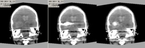
Fig. 5: Left: pre-interventional FDCT reconstruction. Middle: post-coiling reconstruction without MAR. Right: post-coiling reconstruction with MAR applied.
Fig. 5 shows a pre-interventional FDCT and a post coiling FDCT without and with MAR applied,
in order to illustrate the MAR technique. The streaking artifacts have been reduced significantly in the reconstruction with MAR applied.
Soft-tissue structures that are obstructed by the streaking artifacts origining from the coils in the aneurysm sac are clearly visible in the reconstruction with MAR.
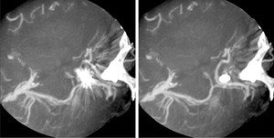
Fig. 6: Axial slab in a contrast enhanced FDCT reconstruction. Left: without MAR. Right: with MAR applied.
In Fig. 6 a reconstruction of a coiled aneurysm is shown,
while the vessels have been filled with diluted contrast medium. The second pass reconstruction (right) shows clearly the vasculature obstructed by the artifacts in the first pass reconstruction (left).
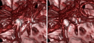
Fig. 7: Zoomed 3D rendering of a contrast enhanced FDCT reconstruction. Left: without MAR. Right: with MAR applied.
Fig. 7 shows a 3D visualization of the reconstruction in Fig. 6.
The streaking artifacts that origin from the coils can form false vessel lumen in the 3D representation (left),
whereas this effect is not present in the MAR corrected image (right).
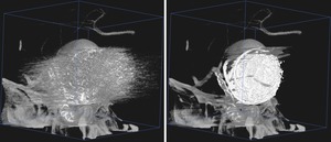
Fig. 8: Contrast enhanced FDCT reconstruction of a stent-assisted coiled large aneurysm. Left: without MAR. Right: with MAR applied.
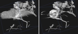
Fig. 9: Contrast enhanced FDCT reconstruction of a stent-assisted coiled aneurysm. Left: without MAR. Right: with MAR applied.
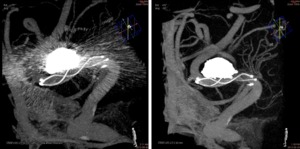
Fig. 10: Contrast enhanced FDCT reconstruction of a stent-assisted coiled aneurysm. Left: without MAR. Right: with MAR applied.
References: Courtesy: Prof. J. Moret, Neuri Hôpital Beaujon, Paris.
Fig. 8, Fig. 9,
and Fig. 10 display volumetric visualizations of stent assisted coiled aneurysms.
The left images show the reconstructions without MAR applied,
and the right images are the result of the MAR algorithm. Though not all stent struts affected by the metal artifacts can be reconstructed in the second pass,
the visibility of the stents has improved considerably.
