There are three major salivary glands,
the parotid,
submandibular and sublingual glands.
Minor salivary glands are diffusely located throughout the aerodigestive tract.
Infectious,
inflammatory,
congenital,
benign and malignant conditions can involve the salivary glands.
Detailed knowledge of the relevant anatomy is essential,
and helps to provide a confident approach to image interpretation.
We propose a guideline for the radiologist in evaluating various lesions based on characteristic imaging features and anatomical relationships.
The parotid space contains the parotid gland which is located superficially and extends over the ascending ramus of the mandible.
It is divided into two compartments; superficial and deep.
The superficial lobe lies beneath the skin and extends superficial to the masseter muscle.
The deep lobe extends deep to the plane of the facial nerve.
Stensen's duct extends anteriorly over the masseter muscle and inserts in the cheek at the second maxillary molar.
The parotid space contains
- parotid gland
- external carotid artery
- retromandilbular vein
- facial nerve
- intra parotid lymph nodes
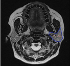
Fig. 1: Axial T2 MRI demonstrating the parotid space containing the parotid gland. A;external carotid artery, V;retromandibular vein; FN;facial nerve.
The floor of the mouth contains the submandibular,
sublingual and minor salivary glands.
The sublingual space is located superomedial to the myelohyoid musle and lateral to the genioglossus geniohyoid complex.
The hyoglossus muscle (an extrinsic muscle of the tongue) extends from the hyoid bone through the sublingual space inserting into the lateral aspect of the tongue.
This separates the lingual artery (lies medial to the hyoglossus muscle) from the lingual vein and whartons duct (submandibular duct) which lie lateral to it.The sublingual glands drain through multiple small ducts that open into the floor of the mouth.
The anterior ducts may join to form a common Bartholin duct.
The sublingual space contains
- sublingual gland
- wharton's duct
- small portion of submandibular gland
- lingual artery and vein
- lingual nerve
- hyoglossus muscle
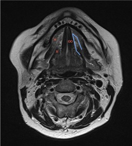
Fig. 2: Axial T2 MRI demonstrating the sublingual space. GG; genioglossus geniohyoid complx, M;Myelohyoid muscle, H; hyoglossus muscle.
The submandibular space is inferior and lateral to the myelohyoid muscle and superior to the fascia lining the platysma muscle.
The ramus of the mandible is lateral to the submandibular space.
The submandibular space contains
- submandibular gland
- submental and submandibular nodal groups
- facial vein and artery
- hypoglossal nerve
- anterior belly of the digastric muscle
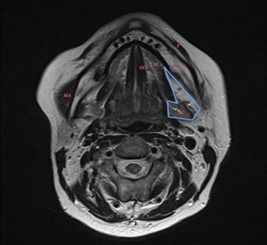
Fig. 3: Axial T2 MRI demonstrating the submandibular space. SG;submandibular gland, GG; genioglossus geniohyoid complex, H;hyoglossus, P;platysma, MA;masseter muscle, M;Myelohyoid muscle, N;submandibular lymph node, RM; ramus of mandible.
There is no posterior fascial border between the sublingual and submandibular spaces allowing communication at the posterior myelohyoid margin.
Similarly no fascial border separates these spaces from the inferior parapharyngeal space allowing for disease infiltration.
Deep parotid space lesions may also infiltrate into the parapharyngeal space displacing the parapharyngeal fat anteriorly.
Other important spaces include:the masticator space,
carotid space,
parapharyngeal space and the pharyngeal mucosal space.
The masticator space contains
- temporalis muscle
- masseter muscle
- medial and lateral pterygoid muscles
- ramus and posterior body of mandible
- mandibular branch of trigeminal nerve
The carotid space contains
- carotid artery
- internal jugular vein
- cranial nerves 9-12
The parapharyngeal space contains
- minor salivary glands
- fat
- internal maxillary artery
- ascending pharyngeal artery
- pterygoid venous plexus
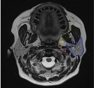
Fig. 4: Axial T2 MRI. PPS; parapharyngeal space, PS; parotid space, MS;masticator space, CS;carotid space.
The pharyngeal mucosal space extends to involve the nasopharynx,
oropharynx and contains
- adenoids (nasopharynx) palatine and lingual tonsils (oropharynx)
- minor salivary glands
- pharyngobasilar fascia
- superior,
middle and inferior constrictor muscles
- levator palatini muscle
- eustachian tube,
torus tubarius
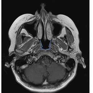
Fig. 5: Axial T1 MRI demonstrating the pharyngeal mucosal space.
