Learning objectives
To present the radiologic anatomy of the normal male urethra in conventional urethrography examinations in association with the type of urethrography performed (either retrograde or voiding) and the age of the patient.
Background
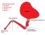
The male urethra extends from the internal urethral sphincter at the neck of the bladder to the external urethral orifice at the tip of the penis.
It is approximately 20cm long (18-25cm) and is divided anatomically and radiologically into two main segments:
the posterior urethra,
which begins at the neck of the bladder,
terminates at the urogenital diaphragm and can be subdivided into prostatic and membranous parts
the anterior urethra,
which extends from the inferior margin of the urogenital diaphragm to the external urethral meatus...
Findings and procedure details
Anterior and posterior normal urethra on RUG and VUG: different methods - different appearance
To understand urethrography images and the differences in the appearance of anterior of posterior urethra depending on the type of the urethrography performed,
one should consider the presence of the external urethral sphincter at the urogenital diaphragm.
The resistance that the external urethral sphincter exerts to the retrograde (ascending) injection of contrast in Retrograde Urethrography (RUG) creates full distension and therefore better depiction of the anterior urethra.
In a similar manner,...
Conclusion
Conventional retrograde and voiding urethrography still remain the best initial imaging methods for the evaluation of urethral anatomy and pathology.
Anterior urethra is visualized on retrograde urethrography and a voiding study is more appropriate for the posterior urethra.In most cases though,
it is necessary to perform both studies in order to ensure that a significant abnormality is not missed out or a normal variant is not misunderstood as pathology,
since the two techniques have complementary roles.
Radiologists should be familiar with normal imaging anatomy of...
Personal information
Radiology Department of "G.Gennimatas" Hospital of Thessaloniki,
GREECE
M.
Arvaniti
D.
Katsiba
D.
Rafailidis
I.
Torounidis
A.
Charsoula
C.
Kaitartzis
C.
Nalmpantidou
A.
Papadimitriou
e-mail address:
[email protected]
mail address:
"G.Gennimatas" Hospital of Thessaloniki
Radiology Department
41 Ethnikis Aminis St.
54635
Thessaloniki
GREECE
References
Kawashima A,
Sandler CM,
Wasserman NF,
LeRoy AJ,
King BF Jr,
Goldman SM.
Imaging of urethral disease: a pictorial review.
Radiographics.
2004 Oct;24 Suppl 1:S195-216.
Kim B,
Kawashima A,
LeRoy AJ.
Imaging of the male urethra.
Semin Ultrasound CT MR.
2007 Aug;28(4):258-73.
Fernbach SK,
Feinstein KA,
Schmidt MB.
Pediatric voiding cystourethrography: a pictorial guide.
Radiographics.
2000 Jan-Feb;20(1):155-68; discussion 168-71.
Levin TL,
Han B,
Little BP.
Congenital anomalies of the male urethra.
Pediatr Radiol.
2007 Sep;37(9):851-62; quiz 945.
Epub 2007 May 22.
Pavlica P,
Barozzi L,...


