This poster is published under an
open license. Please read the
disclaimer for further details.
Type:
Educational Exhibit
Keywords:
Education and training, Normal variants, Cystography / Uretrography, Fluoroscopy, Genital / Reproductive system male, Anatomy
Authors:
A. Charsoula, D. Katsiba, C. Kaitartzis, A. Papadimitriou, C. NALMPANTIDOU, I. Torounidis, D. Rafailidis, M. Arvaniti; Thessaloniki/GR
DOI:
10.1594/ecr2014/C-1524
Background
The male urethra extends from the internal urethral sphincter at the neck of the bladder to the external urethral orifice at the tip of the penis.
It is approximately 20cm long (18-25cm) and is divided anatomically and radiologically into two main segments:
- the posterior urethra,
which begins at the neck of the bladder,
terminates at the urogenital diaphragm and can be subdivided into prostatic and membranous parts
- the anterior urethra,
which extends from the inferior margin of the urogenital diaphragm to the external urethral meatus and can be subdivided into bulbous and penile parts
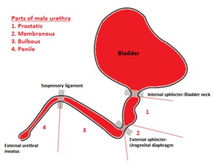
Fig. 1: Parts of male urethra.
1+2: posterior urethra
3+4: anterior urethra
References: John G. Gearhart, Richard C. Rink, Pierre D. E. Mouriquan. Pediatric Urology, 2nd ed. Elsevier Health Sciences, 2009
Conventional urethrography is considered the standard imaging method for studying the morphology and function of the male urethra,
by providing images of the urethral lumen. There are two types of fluoroscopic urethrography: Retrograde Urethrography (RUG) and Voiding Urethrography/Cystourethrography (VUG).
In Retrograde Urethrography (RUG),
contrast media is injected into the external urethral meatus by means of a catheter with the distended balloon positioned in the fossa navicularis or by means of a syringe that occludes the orifice.
This type of urethrography is usually the first one to be performed.
In Voiding urethrography/Cystourethrography (VUG),
images are obtained while the patient urinates in a container.
Filling of the patient’s bladder with contrast is achieved either with the aforementioned retrograde gradual injection of contrast during a preceding RUG or with the insertion of a Foley catheter into the bladder.
VUG is widely used in pediatric radiology in children with a history of urinary tract infection,
as part of a VCUG study.
In both types of urethrography right oblique images are obtained with the patient preferably supine and the penis resting extended along the thigh of the ipsilateral right flexed leg.
This position provides adequate visualization of the whole length of the urethra,
from the bladder neck to the external urethral meatus.

