This poster is published under an
open license. Please read the
disclaimer for further details.
Type:
Educational Exhibit
Keywords:
Neuroradiology brain, MR, CT, Imaging sequences, Blood
Authors:
J. R. Nair1, S. Jaggi2, J. Chankowsky1, C. Torres1, S. H. SHAH2, I. Talwar3, R. del Carpio1; 1Montreal, QC/CA, 2MUMBAI, MAHARASHTRA/IN, 3Mumbai/IN
DOI:
10.1594/ecr2014/C-2095
Background
INTRODUCTION
- Non-traumatic intracranial hemorrhage accounts for approximately 10% to15% of strokes in North America.
- Patients usually present with sudden onset focal neurologic deficit accompanied by headache,
alteration in the level of consciousness,
seizure,
nausea and/or vomiting
- Location:
• Parenchymal (common)
• Subarachnoid
• Subdural
• Intraventricular
• Extradural (rare)
IMAGING MODALITIES:
CT SCAN:
- Plain CT is the initial imaging modality of choice.
- Acute hemorrhage appears hyper dense on plain CT.
MRI:
- Often performed usually after CT to assess cause of hemorrhage
- Sensitive to various stages of hemorrhage.
- GRE/ SWI – most useful sequence.
- MR angiography,
MR venography and MR perfusion may be performed
STAGES OF INTRACEREBRAL HEMORRHAGE:(Fig 1and 2)
STAGE
|
COMPOUND
|
T1W
|
T2W
|
HYPERACUTE
(0-FEW HRS)
|
OXYHEMOGLOBIN
|
ISOINTENSE
|
HYPERINTENSE
|
ACUTE
(FEW HRS–
3 DAYS
|
DEOXYHEMOGLOBIN
|
ISO-HYPOINTENSE
|
HYPOINTENSE
|
SUB-ACUTE
(3DAYS–3 WEEKS)
|
METHEMOGLOBIN
- EARLY
-- LATE |
HYPERINTENSE
HYPERINTENSE
|
HYPOINTENSE
HYPERINTENSE
|
CHRONIC
(MORE THAN 3 WEEKS)
|
HAEMOSIDERIN
|
HYPOINTENSE
|
HYPOINTENSE
|
Fig. 2: MRI- Stages of Intracerebral Hemorrhage
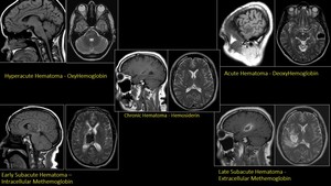
ETIOLOGIES:
•Hypertension
•Cerebral Amyloid Angiopathy
•Hemorrhagic
transformation of ischemic stroke
•Aneurysmal subarachnoid hemorrhage
•Vascular malformation
•Venous Thrombosis
•Neoplasia – Primary,
Metastases
|
•Vasculitis,Vasculopathy,
Vasospasm
•Subdural Hemorrhage
•Anticoagulation
•Clotting factor deficiency
•Hepatic dysfunction
•Metabolic disorders
•Viral encephalitis
|
CAUSES OF NON-TRAUMATIC INTRACEREBRAL HEMORRHAGE