Learning objectives
The purpose of this educational exhibit is to: illustrate the specific changes in MRI patterns of the brain in patients with сlinically confirmed diagnosis of CerebralAutosomal Dominant Arteriopathy with Sub-cortical Infarcts and Leukoencephalopathy (CADASIL).
Background
Cerebral Autosomal Dominant Arteriopathy with Subcortical Infarcts and Leukoencephalopathy (CADASIL) is an inherited autosomal dominant vascular dementiacaused by mutation in the Notch3 gene of chromosome 19,
alters the muscular walls of small blood vessels in the white matter and basal ganglia of the brain.
Thisresults to progressive symptoms of transient ischemic attacks,
strokes,
and vascular dementia.
The most common clinical manifestations usually occur between 40and 50 years of age,
although MRI is able to detect signs of the disease years prior to clinical manifestation of...
Findings and procedure details
We use 1.5T MRI (Magnetom Essenza,
Siemens AG,
Medical Solutions,
Magnetic Resonance,
Henkestr.
127,
Erlangen,
Germany) with protocol includes T1/T2-weighted,
T2-FLAIR,
DWI and SWI sequences.
Contrast enhancement is usually not required.
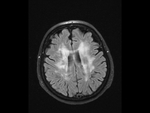
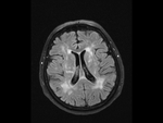
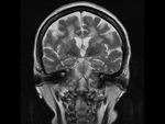
MRI findings includes widespread confluent white matter hyperintensities,
predominantly in the frontal and temporal lobes,
multiple hyperintence lesions in thebasal ganglia,
thalamus and pons,
reduction volume of the brain.
Cerebral microhaemorrhages are also often findings without a characteristic distribution.
They were defined as focal areas of signal loss on T2-weighted spin-echo images that were...
Conclusion
Patients with lacunar infarcts in particular should be monitored closely becauselacunar infarcts are associated with disability and cognitive decline.
MRI of the Brain is the no-invasive investigation of choice in patients with suspect CADASIL disease,
allow to accurately assess the prevalence and dynamic ofpathological changes.
Personal information
Alexey Kokunin,
M.D.
Regional Diagnostic Center,
MRI Department(Nizhniy Novgorod,
Russia)
Balakhna Central Regional Hhospital,Chief of Radiology Department(Balakhna,
Russia)
[email protected]
References
1) Ruchoux MM,
Guerouaou D,
VandenhauteB,
Pruvo JP,
Vermersch P,
Leys D.
SystemicNEURORADIOLOGY:Progression of MR Abnormalities in CADASIL Liem et al970 Radiology: Volume 249: Number 3—December 2008 vascular smooth muscle cell impairment incerebral autosomal dominant arteriopathywith subcortical infarcts and leukoencephalopathy.
Acta Neuropathol 1995;89(6):500 –512.
2) Liem MK,
van der Grond J,
Haan J,
et al.
Lacunar infarcts are the main correlate with cognitive dysfunction in CADASIL.
Stroke 2007;38(3):923–928.
3) Viswanathan A,
Gschwendtner A,
Guichard JP,
et al.
Lacunar lesions are independently associated with disability and...


