This poster was previously presented at the 69th Korean Congress of Radiology (KCR 2013) in Seoul.
This poster is published under an
open license. Please read the
disclaimer for further details.
Keywords:
CT-Angiography, Vascular, Cardiovascular system, Cardiac, eLearning, Acute
Authors:
E. Lee1, S. I. Choi2, E. J. Chun2, Y. K. Kim3; 1Seongnam-si, Gyeonggi-do/KR, 2Seongnam-Si/KR, 3Seoul/KR
DOI:
10.1594/ecr2014/C-2377
Aims and objectives
1.
Introduce the causes of variant angina
2.
Understand the clinical and angiographic manifestation of variant angina
3.
Show the potential of cardiac MDCT imaging to verify variant angina on the basis of our cardiac MDCT registry with 58,
372 cases.
: This exhibit aims to illustrate various imaging findings of several subtypes of variant angina
Introduction>
•Angina pectoris is caused by transient myocardial ischemia due to an imbalance between myocardial oxygen demand and supply.•Classical or stable angina is characterized by (1) the attack is induced by exertion and relieved by rest or nitroglycerin administration,
and (2) the attack is associated with transient ST segment depression in the electrocardiogram.•This form of angina has been well known for more than 200 years since its description by Heberden.•In 1959,
Prinzmetal et al described a new form of angina pectoris which differed sharply from the classical angina and named it ‘‘variant form of angina pectoris.’’
Diagnosis of variant angina>
•coronary vasospastic angina (VA) denotes chest discomfort with angina pectoris that occurred at rest,
usually at night,
or during the early morning.•The pain is often associated with a transient elevation of the ST segment greater than 2 mm on the electrocardiogram .
Its cause is well known as vasospasm,
a narrowing of the coronary arteries caused by the contraction of smooth muscle tissue in the vessel walls.•Vasospasm is confirmed by conventional coronary angiography with provocation using acetylcholine or ergonovine.
Coronary angiographic changes>
•coronary spasm appears in angiographically normal arteries as well as those with organic stenosis.
One study reported that 70% of variant angina had normal or near normal coronary arteries.•Spasm may be diffuse or diffuse plus focal and may even migrate from site to site.
Spasm occurs not only at one large coronary artery but also at two or three large arteries separately or simultaneously in the same patients.
Multi-vessel spasm was demonstrated in 52% of the patients.• The patients with multi-vessel coronary spasm have the following characteristics: (1) most of them have angiographically normal coronary arteries; (2) they are resistant to treatment and often require larger amounts of CCBs to suppress the attacks,
which often recur on cessation of the drugs; (3) they are more likely to have lethal arrhythmias such as ventricular tachycardia or ventricular fibrillation and are more likely to suffer from sudden death.
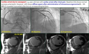
Fig. 6

