ECR 2015 / C-0087
Computed Tomographic assessment of Body Trauma with a Triple Fractionated Intravenous Contrast Injection Protocol
This poster is published under an open license. Please read the disclaimer for further details.
Congress:
ECR 2015
Poster Number:
C-0087
Type:
Scientific Exhibit
Keywords:
CT, Trauma, Emergency, Abdomen, Contrast agent-intravenous, Radiation safety
Authors:
R. Lakshmanan1, R. M. Mendelson2, S. Rao2, P. D. Fatovich2; 1Perth, Western Australia/AU, 2Perth/AU
DOI:
10.1594/ecr2015/C-0087

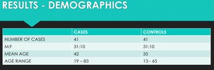
Fig. 5:
Demographics table

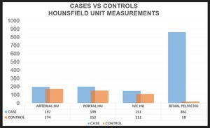
Fig. 6:
Comparison of HU measures between the cases and controls

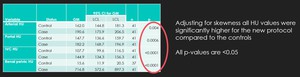
Fig. 7:
Statistical significance of the higher HU values in the case group.
. References: Royal Perth Hospital, Radiology Department")
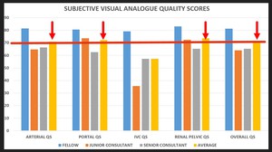
Fig. 8:
Average subjective quality scores for each reviewer and across all reviewers...

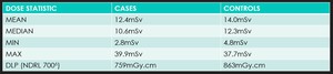
Fig. 9:
Radiation dose statistics

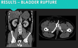
Fig. 10:
Detection of extraperitoneal bladder rupture on the initial scan due to the...

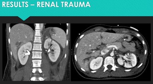
Fig. 11:
Patient with high grade renal trauma showing no evidence of collecting system...