This poster is published under an
open license. Please read the
disclaimer for further details.
Keywords:
CT, Trauma, Emergency, Abdomen, Contrast agent-intravenous, Radiation safety
Authors:
R. Lakshmanan1, R. M. Mendelson2, S. Rao2, P. D. Fatovich2; 1Perth, Western Australia/AU, 2Perth/AU
DOI:
10.1594/ecr2015/C-0087
Aims and objectives
Background
It is well established that CT in body trauma is associated with improved patient survival1,
it is therefore an indispensible tool in the evaluation of the trauma patient.
The benefits of CT in trauma are mitigated by the high cumulative radiation dose experienced by a patient during their admission,
for which CT is the main culprit2.
Therefore by optimizing the dose for trauma CT patients can still enjoy the survival benefit while reducing the long term potential radiation related harm.
The Problem
The high cumulative radiation dose in trauma patients occurs in generally a young demographic.
Additionally,
many patients presenting with abdominal trauma warrant exclusion of urinary tract injuries through the use of a delayed CT and CT cystogram,
this further contributes to radiation burden.
Patients who undergo delayed scanning for pyelographic assessment will spend in excess of 10 minutes on the scanning table,
this leaves a potentially unstable trauma patient extremely vulnerable whilst on the scanner, where there is limited space and resources to perform acute life support.
Aims and Objectives
We aim to show that through the use of our novel triple fractionated contrast injection technique,
good quality images can be obtained whilst reducing radiation dose,
reducing the time a potentially unstable patient spends on the CT table and assisting in the early exclusion of clinically significant renal collecting system injuries.
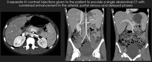
Fig. 1: Example case from our study demonstrating good arterial, portal venous and pyelographic enhancement.
References: Royal Perth Hospital, Radiology Department

