This poster is published under an
open license. Please read the
disclaimer for further details.
Type:
Educational Exhibit
Keywords:
Education and training, Normal variants, MR, Conventional radiography, Paediatric, Musculoskeletal joint
Authors:
A. Lakatos, B. Lombay; Miskolc/HU
DOI:
10.1594/ecr2015/C-0204
Background
Blount-disease is characterized by disorganized enchondral ossification (osteochondrosis) of the medial portion of the epiphyseal and metaphyseal areas of the proximal tibia [1].
The exact etiology is unknown,
but there is strong association with childhood obesity,
which suggests a mechanical background.
There is an inbalance between the nutritional needs of the growing bone [2,
3,
4].
Risk factors for Blount-disease:
- ethnicity or race - higher incidence in children of African origin
- gender - more common in females
- genetics - autosomal dominant and recessive form
- early walking - less than one year of age
- mechanical stress
- obesity
Based on the onset of the clinical findings two distinct – the early and late onset - forms are recognized [1] (Fig.
1).
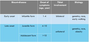
Fig. 1: Types of Blount-disease.
The early onset or infantile Blount-disease,
occurring before the age of 4 years,
usually involves the tibia bilaterally.
The late onset Blount-disease is further subcategorized into juvenile (between the age of 4-10 years) and adolescent form (beginning after the age of 10 years).
These latter two subtypes mainly involve the tibia unilaterally.
If remains unresolved,
Blount-disease can lead to progressive multiplanar deformities in the lower extremity as:
- tibial varus,
- genu procurvatum and
- internal torsion with
- leg length discrepancy resulting early osteoarthritis of the knee [5].
With accurate radiographic evaluation and orthopedic treatment these complications are partially or completely avertable.
