CT and MRI provide detailed anatomy of the skull base.
Both are complementary techniques.
CT is an excellent modality for delineation of the bone structures and MRI provides superior soft tissue contrast it is better to evaluate the intracranial extension of the lesions.
The sagittal plane provides a good vision of the skull base to decide the best surgical approach.
It is important to be aware of the contraindications to surgery that include,
invasion of the cavernous sinuses,
optic nerves and chiasm.
ASSESSMENT OF LESION EXTENSION
CHECKLIST
- Location and extention of the lesion
- Amount of bony skull base involvement
- Intracranial and orbital invasion
- Cavernous sinus invasion
- Cranial nerve and vessel involvement
- Dural invasion
- Optic nerve or quiasm invasion
ANTERIOR SKULL BASE LESIONS
Anterior skull base defects
- Ethmoid meningoencephalocele
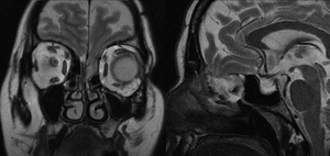
Fig. 9: Meningoencephalocele
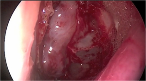
Fig. 10: Close up view of surgery of the meningoencephalocele
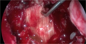
Fig. 11: Repair and closure of the anterior skull defect with fascia lata
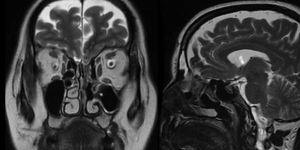
Fig. 12: Repaired meningoencephalocele
Ethmoid sinuses infections
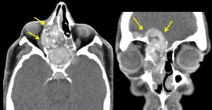
Fig. 19: Fungal rhinosinusitis with orbital and cranial invasion (yellow arrows)
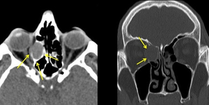
Fig. 20: Right ethmoidal mucocele with orbital invasion (yellow arrows)
Esthesioneuroblastoma
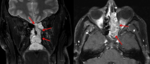
Fig. 15: Esthesioneuroblastoma arising from the olfatry groove and invading the ethmoid sinuses (red arrows) and extending into the nasopharynx
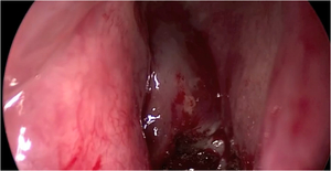
Fig. 16: Close up view of the esthesioneuroblastoma at surgery
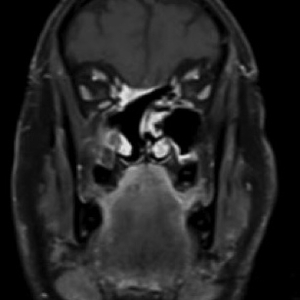
Fig. 17: Follow up MRI one year after surgery. Surgical changes with linear enhancement in the ethmoid region extending to anterior skull base
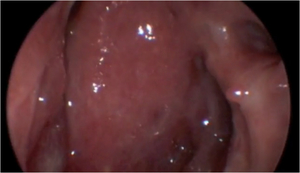
Fig. 18: Endoscopic image of the surgical cavity. The mucosa is vascularized and shows a normal appearance
Ethmoid sinuses neoplasms
- Benign (osteoma,
fibrous dysplasia,
meningioma)
- Malignant (squamous cell,
adenocarcinoma,
lymphoma,
undifferentiated carcinoma,
sarcoma)
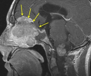
Fig. 21: Meningioma of the olfatory groove. Large mass in the anterior cranial fossa, pushing the brain superiorly, invading the ethmoid sinuses below and extending to the planum sphenoidale posteriorly (yellow arrows)
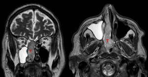
Fig. 13: Sinonasal adenocarcinoma involving the right ethmoid sinuses and nasal fossa. The anterior skull base is not invaded
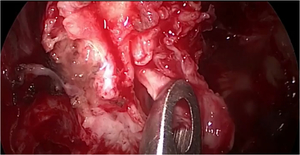
Fig. 14: Close up view of the adenocarcioma at surgery
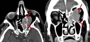
Fig. 22: Lymphma involving the left ethmoidal cells (asterisk) infiltrating the lamina papiracea and extending into the orbit (red arrows)
IMAGING ASSESMENT OF ASB INVASION
Invasion of the anterior cranial fossa is better depicted by MRI
When the tumor extends to de anterior cranial fossa three different situations can be seen on imaging:
- The neoplasm contacts the ASB but there is not invasion
- The neoplasm encroaches the cribriform plate,
the dura is thickened but not invaded (linear thickening <5mm and enhancement)
- The neoplasm encroaches the dura (dural thickening >5mm,
nodular enhancement,
pial enhancement)
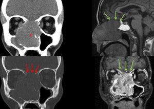
Fig. 6: Ethmoid sinuses squamous cell carcinoma
CT demostrates the soft tissue mass involving and destroying the sinuses, with orbital invasion. The bone window shows destruction of the cribiform plate (red arrows)
MRI shows the mass with no dural thickening or enhancement (green arrows). MRI is superior to CT to rule out dural or brain invasion
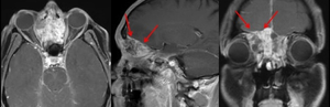
Fig. 7: Intestinal-type sinonasal adenocarcinoma.
MRI shows extension of the tumor to the anterior skull base. The neoplasm encroaches the cribriform plate, T1W gadolium enhanced MRI depicts the linear dural thickening (<5mmm) and enhancement (red arrows). The dura is thickened but not invaded
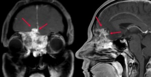
Fig. 8: Intestinal-type adenocarcinoma
MRI shows the neoplasm encroaching and invading the dura. The dura is thickened (>5mm). T1W MRI with gadolinium depicts the nodular dural and pial enhancement(red arrows)
Midline Sagittal CSB LESIONS
Originate from the greater sphenoid wing,
cavernous sinus,
cranial nerves and petroclival synchondrosis
- Sphenoid body,
sphenoid sinus (infections,
neoplasms)
- Clivus (metastasis,plasmacytoma,chordoma,chondrosarcoma)
- Sella turcica (macroadenoma,
meningioma,
craniopharyngioma)
- Nasopharynx (infections,
neoplasms)
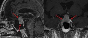
Fig. 23: Macroadenoma with suprasellar extension (red arrows)
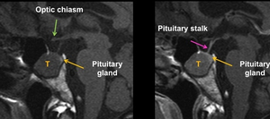
Fig. 24: Intrasphenoid macroadenoma.The tumor originates in the pituitary gland with intrasphenoid growth
Fig. 25: Intrasellar and suprasellar lung metastasis

Fig. 26: Craniopharyngioma. Heterogeneous midline suprasellar mass with solid and cystic components.The midline, the pituitary gland is displaced downward
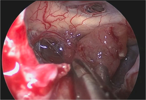
Fig. 27: Close up view of the clival chordoma surgery
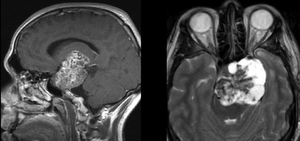
Fig. 28: Clival chordoma. Midline soft tissue mass with cystic and low signal intensity areas, with a characteristic pop corn type of enhancement
Parasagittal CSB LESIONS
Originate from the greater sphenoid wing,
cavernous sinus,
cranial nerves and petroclival synchondrosis
- Developmental lesions ( trans-sphenoidal cephaloceles)
- Cavernous sinus lesions (primary and secondary cranial nerve tumors,
vascular lesions,
meningiomas and inflammatory conditions)
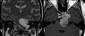
Fig. 29: Chordoma arising at the petroclival junction. Parasagittal central skull bilobulated enhancing mass
Lateral CSB LESIONS
The lateral CSB includes the lateral aspect of the greater sphenoid wings,
the lateral aspect of the temporal bone and the TMJ. It is a crossroads between the orbit,
the middle cranial fossa and temporal fossa
- Meningiomas
- Osteosarcomas
- Metastasis
- Lymphoma
- Synovial chondromatosis (TMJ)
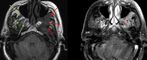
Fig. 30: Masticator space lymphoma. Infiltrating process on the left masticator space (red arrows),normal masticator space on the right (green arrows)
