ECR 2015 / C-0264
Imaging of the anterior and central skull base as a guide for endoscopic skull surgery
This poster is published under an open license. Please read the disclaimer for further details.
Congress:
ECR 2015
Poster Number:
C-0264
Type:
Educational Exhibit
Keywords:
Head and neck, Anatomy, MR, CT, Diagnostic procedure, Education, Image registration, Pathology, Image verification
Authors:
L. Oleaga Zufiría, I. Alobid, J. Berenguer, I. Valduvieco, E. Verger; Barcelona/ES
DOI:
10.1594/ecr2015/C-0264

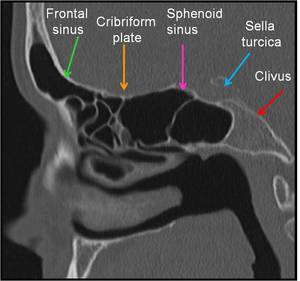
Fig. 1:
Sagittal CT image. Anatomic landmarks for endoscopic skull surgery

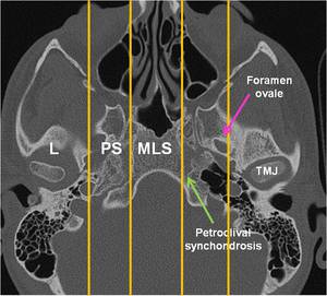
Fig. 4:
Axial CT demonstrating the anatomic landmarks in central skull base

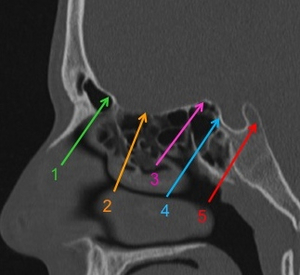
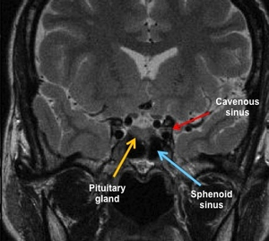
Fig. 3:
Sagittal CT image with showing the entry points of surgical corridors for...