This poster is published under an
open license. Please read the
disclaimer for further details.
Keywords:
Breast, Molecular imaging, Oncology, Ultrasound, Diagnostic procedure, Physiological studies, Blood, Cancer, Tissue characterisation
Authors:
R. Butler1, L. F. Tucker2, P. Lavin3, M. J. Ulissey4, A. T. Stavros4; 1New Haven, CT/US, 2Wirtz, VA/US, 3Framingham, MA/US, 4San Antonio, TX/US
DOI:
10.1594/ecr2015/C-1046
Aims and objectives
Opto-acoustic (OA) Imagio® (Seno Medical Instruments,
San Antonio,
TX) is a novel investigational device (Figure 1) with functional modality that integrates laser optics with ultrasound in an effort to improve the performance of currently available breast imaging modalities without the use of IV contrast or ionizing radiation.
A fusion of anatomic and functional modalities,
OA imaging provides both conventional B-mode images and co-registered real time color maps that demonstrate tumor vascularity and the relative amount of hemoglobin oxygenation within and around breast tumors.
The technology is based on the observation that malignant tumors must generate neovessels to grow larger than 2 mm and are also more metabolically active,
extracting oxygen from hemoglobin to a greater degree than benign neoplasms or normal tissue [1,
2].
This added information may help differentiate benign from malignant masses even when their gray-scale sonographic morphologic features overlap.
In contrast to conventional ultrasound,
which both emits and receives high-frequency sound waves,
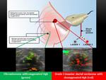
OA imaging emits short pulses of laser light at two wavelengths that correspond to the absorption peaks of oxygenated (1064 nm) and deoxygenated (757 nm) hemoglobin.
These very brief bursts of low energy laser cause a momentary heating and expansion of blood,
which is detected and color-coded as green for oxygenated hemoglobin and red for deoxygenated hemoglobin.
The color-coded,
opto-acoustic data is co-registered with the gray-scale ultrasound image in real time (Figure 2).
In this study,
we correlated OA imaging findings with histopathologic features of breast cancers to elucidate the histopathologic basis for the findings of this new functional imaging platform.


