ECR 2015 / C-1578
The palmar aponeurosis: dynamic evaluation with 22 Mhz high-resolution ultrasound (HR-US)
This poster is published under an open license. Please read the disclaimer for further details.
Congress:
ECR 2015
Poster Number:
C-1578
Type:
Educational Exhibit
Keywords:
Image verification, Education, Ultrasound, Musculoskeletal system
Authors:
S. Perugin Bernardi, A. Arcidiacono, A. Corazza, R. Sartoris, A. Muda, E. Silvestri; Genoa/IT
DOI:
10.1594/ecr2015/C-1578
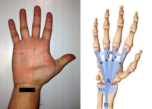
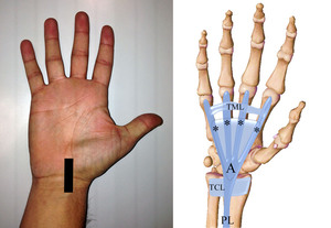
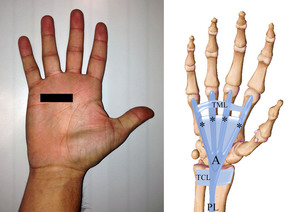
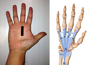
 and palmar aponeurosis (A). TML, transverse metacarpal ligament; TCL, transverse carpal ligament; dashed triangle, central bundle; *, slip for the fingers.")
Fig. 1:
Anatomical scheme of palmari longus (PL) and palmar aponeurosis (A). TML,...

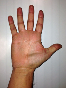
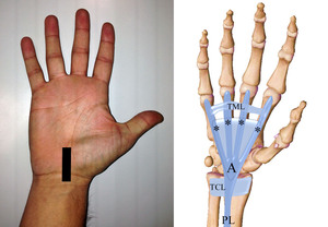
Fig. 2:
Position of the hand to evaluate the palmar aponeurosis.
 tendon on axial plane, proximal to carpal tunnel, correlated with anatomical scheme of PL and palmar aponeurosis (A). TML, transverse metacarpal ligament; TCL, transverse carpal ligament; dashed triangle, central bundle; *, slip for the fingers.")
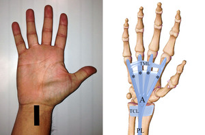
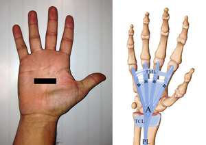
Fig. 3:
Probe position to evaluate palmaris longus (PL) tendon on axial plane, proximal...
, proximal to carpal tunnel.")
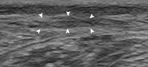
Fig. 4:
Axial scan of palmaris longus tendon (arrowheads), proximal to carpal tunnel.
 tendon with longitudinal scan proximal to carpal tunnel correlated with anatomical scheme of PL and palmar aponeurosis (A). TML, transverse metacarpal ligament; TCL, transverse carpal ligament; dashed triangle, central bundle; *, slip for the fingers.")
Fig. 5:
Probe position to evaluate palmaris longus (PL) tendon with longitudinal scan...
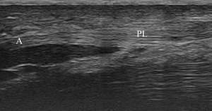
, proximal to carpal tunnel; fl, flexor tendon.")
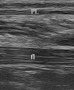
Fig. 6:
Longitudinal scan of palmaris longus tendon (PL), proximal to carpal tunnel;...
 tendon on axial plane at level of carpal tunnel, correlated with anatomical scheme of PL and palmar aponeurosis.")
Fig. 7:
Probe position to evaluate palmaris longus (PL) tendon on axial plane at level...
, at the level of the carpal tunnel.")
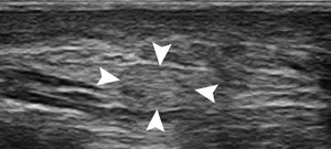
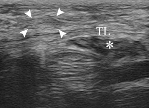
Fig. 8:
Axial scan of palmaris longus tendon (arrowheads), at the level of the carpal...
 tendon with longitudinal scan at level of carpal tunnel, correlated with anatomical scheme of PL and palmar aponeurosis (A). TML, transverse metacarpal ligament; TCL, transverse carpal ligament; dashed triangle, central bundle; *, slip for the fingers.")
Fig. 9:
Probe position to evaluate palmaris longus (PL) tendon with longitudinal scan...
, at the level of the carpal tunnel. Note the ralationship with median nerve (*). fl, flexor tendons.")
Fig. 10:
Longitudinal scan of palmaris longus tendon (PL), at the level of the carpal...
 tendon with transverse carpal ligament, correlated with anatomical scheme of PL and palmar aponeurosis (A). TML, transverse metacarpal ligament; TCL, transverse carpal ligament; dashed triangle, central bundle; *, slip for the fingers.")
Fig. 11:
Probe position to evaluate the relationship of palmaris longus (PL) tendon...
, at the level of the carpal tunnel. *, median nerve; TL, transverse carpal ligament.")
Fig. 12:
Axial scan of palmaris longus tendon (arrowheads), at the level of the carpal...
 tendon which continues in palmar aponeurosis (A), correlated with anatomical scheme of PL and palmar aponeurosis (A). TML, transverse metacarpal ligament; TCL, transverse carpal ligament; dashed triangle, central bundle; *, slip for the fingers.")
Fig. 13:
Probe position to evaluate palmaris longus (PL) tendon which continues in...
 when it continues in the palmar aponeurosis (A).")
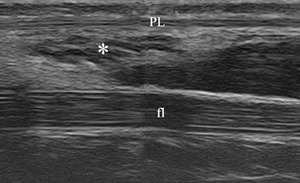
Fig. 14:
Longitudinal scan of palmaris longus tendon (PL) when it continues in the...
 and palmar aponeurosis (A).TML, transverse metacarpal ligament; TCL, transverse carpal ligament; dashed triangle, central bundle; *, slip for the fingers.")
Fig. 15:
Probe position to evaluate the central bundle of palmaris aponeurosis...
 at middle of the palm.; fl, flexor tendons.")
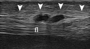
Fig. 16:
Axial scan of the central bundle of palmar aponeurosis (arrowheads) at middle...
 at middle of the palm.; fl, flexor tendons. Doppler sign on the vassels of the superficial palmer arch.")
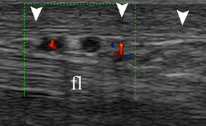
Fig. 17:
Axial scan of the central bundle of palmar aponeurosis (arrowheads) at middle...
 and palmar aponeurosis (A).TML, transverse metacarpal ligament; TCL, transverse carpal ligament; dashed triangle, central bundle; *, slip for the fingers.")
Fig. 18:
Probe position to evaluate with axial scan the finger slips of central bundle...
 for the IV finger¸a, artery of the superficial palmer arch.")
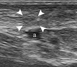
Fig. 19:
Axial scan of the finger slip of palmar aponeurosis (arrowheads) for the IV...
 and palmar aponeurosis (A). TML, transverse metacarpal ligament; TCL, transverse carpal ligament; dashed triangle, central bundle; *, slip for the fingers.")
Fig. 20:
Probe position to evaluate with longitudinal scan the finger slips of central...
 for the III finger¸flIII, flexor tendon of III finger.")
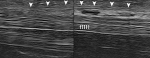
Fig. 21:
Longitudinal scan of the finger slip of palmar aponeurosis (arrowheads) for the...
 for the III finger¸flIII, flexor tendon of III finger.")
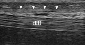
Fig. 22:
Longitudinal scan of the finger slip of palmar aponeurosis (arrowheads) for the...