712 patients presented 774 fractures of the temporal bone.
6.5% (n=46) of the patients had bilateral temporal bone fractures.
External ear fractures were found in 50% of the cases (n=320),
mostly concerning the anterior wall (75%,
n=240),
but external ear fractures were isolated in only 4% (n=31) of the cases.
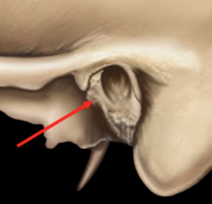
Fig. 1: External ear fractures, 50% of all fractures (arrow)
References: Department of Radiology 1, Strasbourg University Hospital, Strasbourg University, France 2015
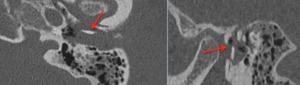
Fig. 2: External ear fracture on an axial plane and sagittal plane : same patient (red arrows)
References: Department of Radiology 1, Strasbourg University Hospital, Strasbourg University, France 2015
Middle ear fractures were found in 79% (n=611) of the cases.
352 of these fractures were reanalyzed and divided in 6 types,
from A to F,
according to the entry point of the fracture in the horizontal part of the squamous bone and/or the petrous bone.
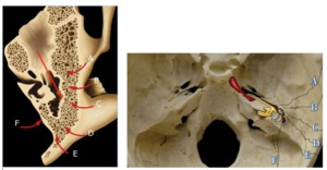
Fig. 3: Middle ear fractures, classified from type A to type F.
References: Department of Radiology 1, Strasbourg University Hospital, Strasbourg University, France 2015
16% (n=56) were type A fractures,
with an entry point anterior to the external auditory meatus (EAM),
in the anterior part of the horizontal squamous bone (squamous part of the temporomandibular joint).
In 50% of the cases,
type A fractures were associated with other fracture types with different entry points.
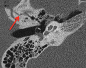
Fig. 4: Type A fracture (non-enhanced CT, axial plane) of the anterior part of the horizontal squamous bone (red arrow)
References: Department of Radiology 1, Strasbourg University Hospital, Strasbourg University, France 2015
27% (n=95) were type B fractures with an entry point through the roof of the EAM,
in the middle part of the horizontal squamous bone.
There was always a fracture of the wall of the attic,
which is the internal part of the squamous bone.
An association with other type of fractures was found in one third of the cases.
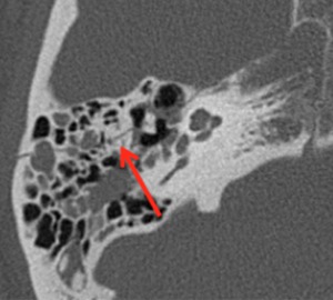
Fig. 5: Type B fracture (non-enhanced CT, axial plane) of the middle part of the horizontal squamous bone -roof of the external auditory meatus- (red arrow)
References: Department of Radiology 1, Strasbourg University Hospital, Strasbourg University, France 2015
Type C fractures were the most common representing 40% of middle ear fractures (n=143),
with an entry point located behind the EAM,
at the level of the lateral wall of the antrum.
An association with other type of fractures was found in one third of the cases.
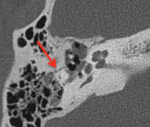
Fig. 6: Type C fracture (red arrow), the most common type. Non-enhanced CT, axial plane.
References: Department of Radiology 1, Strasbourg University Hospital, Strasbourg University, France 2015
Type D fractures were less common (6%,
n=20) with an entry point through the superior-posterior petrosquamous fissure.
It entered the temporal bone through the petrosquamous V: the inner part of the V was made of the petrous bone and the external part was made of squamous bone,
the junction between the two giving the petrosquamous fissure.
It then coursed in the antrum before following the orientation of the tympanic cavity.
An association with other type of fractures was found in one third of the cases.
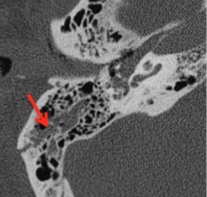
Fig. 7: Type D fracture (red arrow), non-enhanced CT, axial plane
References: Department of Radiology 1, Strasbourg University Hospital, Strasbourg University, France 2015
Type E fractures had an entry point at the posterior part of the petrous bone (internal branch of the petrosquamous V),
close to the lateral sinus (6%,
n=20).
Then the fracture ran between petrous and squamous bone,
following the orientation of the tympanic cavity.
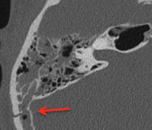
Fig. 8: Type E fracture (red arrow), non-enhanced CT, axial plane
References: Department of Radiology 1, Strasbourg University Hospital, Strasbourg University, France 2015
Type F fractures ran through the medial wall of the petrous bone in 5% of the cases (n=18).
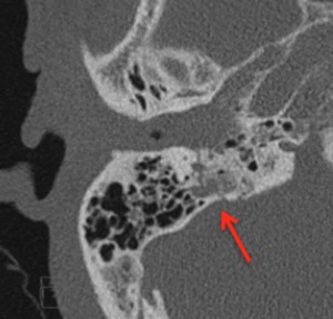
Fig. 9: Type F fracture (red arrow), non-enhanced CT, axial plane
References: Department of Radiology 1, Strasbourg University Hospital, Strasbourg University, France 2015
Isolated inner ear fractures represented 8% (n=63) of all fractures.
These fractures were more difficult to see than middle ear ones as they were very thin in a very compact bone.
All these fractures had an entry point through the internal and posterior wall of the petrous bone,
perpendicular to its axis in the vast majority of cases.
51 of these were reanalyzed.
The cochlea was involved in 55% (n=28),
sometimes associated with an internal auditory canal fracture (which is never isolated).
Fracture of the vestibule were more frequent,
occurring in 73% of the cases (n=37) and rarely isolated (only in 16% of the cases,
n=8).
Semicircular canals fractures were also frequent: 49% of the cases (n=26) whether isolated or not.
Because inner ear fractures were mostly perpendicular,
they had a tendency to reach the oval (49%,
n=26,
especially in its posterior part) and round windows (59%,
n=30),
an enlarged CT image being useful for their study.
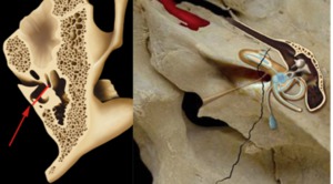
Fig. 10: Isolated inner ear fracture (red arrow), entry point at the internal part of the petrous bone. 8% of the cases.
References: Department of Radiology 1, Strasbourg University Hospital, Strasbourg University, France 2015
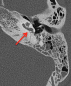
Fig. 11: Isolated inner ear fracture through the cochlea (red arrow). Non enhanced CT, axial plane
References: Department of Radiology 1, Strasbourg University Hospital, Strasbourg University, France 2015
An association of inner and middle ear fractures represented 5% of the fractures (n=39).
In most cases,
they were transversal,
first concerning the middle ear then reaching the inner ear.
In one fourth of the cases it was the contrary,
with fractures first coming from the internal part of the petrous bone and then coursing in the tympanic cavity.
Longitudinal fractures of the middle and inner ear were very rare,
1% of the cases (n=7).
36 cases of associated middle and inner ear fractures were reanalyzed: 22% (n=8) reached the internal auditory canal,
44% (n=16) the cochlea,
53% (n=19) the vestibule and 61% (n=22) the semi-circular canals (either associated to other fractures or isolated).
In this series,
we found 36% of oval window fractures (n=13) and 42% of round window fractures (n=15).
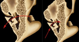
Fig. 12: Associated inner and middle ear fracture in 5% of cases (red arrow). (a) Fracture line going through the middle ear to the inner ear (3/4 of cases). (b) Fracture line going through the inner ear to the middle ear (1/4 of cases).
References: Department of Radiology 1, Strasbourg University Hospital, Strasbourg University, France 2015
All the fractures don’t involve the cavities.
In 2% of the fractures ran outside the temporal bone cavities.
