We propose an easy way of reading of middle ear compartements on MRI images without the need of CT/ MR superposition.
MRI protocol :
- axial images are directly acquired in a plane parallel to the orbital roof.
This plane is parallel to the lateral semicircular canal.
- coronal sections are perpendicukar to the axial ones.
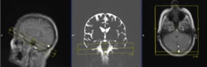
Fig. 1: MRI protocol
References: Department of Radiology 1, Strasbourg University Hospital, Strasbourg University, France 2015
The middle ear is divided in three different compartments on the coronal section: From the top to the bottom:
- Epitympanum (E)
- Mesotympanum (M)
- Hypotympanum (H)
The Epitympanum (E,
yellow) is located above the level of the external auditory canal (EAC).
Notice that the roof of the EAC is located at the same level as the floor of the internal auditory canal.
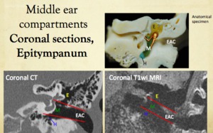
Fig. 2: Coronal sections (dry temporal bone, coronal CT, coronal T1wi MRI), Epitympanum
References: Department of Radiology 1, Strasbourg University Hospital, Strasbourg University, France 2015
The lower Epitympanum (e) is located laterally to the ampulla and the convexity of the lateral semicircular canal (LSCC) and above.
The antrum (a) is posterior to a line (red line) perpendicular to the posterior semicircular canal (PSCC).
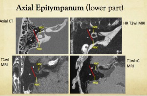
Fig. 3: Axial Epitympanum (lower part), axial CT, axial HRT2wi MRI, axial T1wi MRI, axial T1+C wi MRI
References: Department of Radiology 1, Strasbourg University Hospital, Strasbourg University, France 2015
The upper Epitympanum (e) is located above the level of the lateral semicircular canal,
in front of a line that is perpendicular to the posterior semicircular canal (red line).
The antrum (a) is posterior to a line (red line) perpendicular to the posterior semicircular (PSCC).
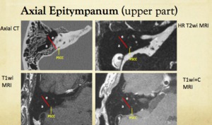
Fig. 4: Axial Epitympanum (lower part), axial CT, axial HRT2wi MRI, axial T1wi MRI, axial T1+C wi MRI
References: Department of Radiology 1, Strasbourg University Hospital, Strasbourg University, France 2015
The Mesotympanum (M,
green) is located between the promontory and the tympanic membrane,
in front of the external auditory meatus.
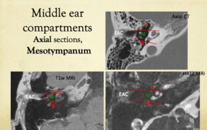
Fig. 5: Coronal sections (dry temporal bone, coronal CT, coronal T1wi MRI), Mesotympanum
References: Department of Radiology 1, Strasbourg University Hospital, Strasbourg University, France 2015
The Mesotympanum (m) is located between the promontory (pr) and the tympanic membrane,
facing the external auditory canal (EAC).
Medially it is facing the basal turn of the cochlea (bt) and the inferior part of the vestibule.
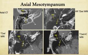
Fig. 7: Axial Mesotympanum (axial CT, axial T2wi MRI, axial T1wi MRI, axial T1+C wi MRI)
References: Department of Radiology 1, Strasbourg University Hospital, Strasbourg University, France 2015
The Mesotympanum is the compartment where the round window (RW,
at the posterior extremity of the basal turn of the cochlea) and the oval windows (OW,
which opens at the inferior and lateral part of the vestibule) are located.

Fig. 6: Round window and oval window levels (axial CT, axial T1wi MRI, axial HRT2wi MRI)
References: Department of Radiology 1, Strasbourg University Hospital, Strasbourg University, France 2015
The Hypotympanum (H,
blue) is located below the level of the floor of the external auditory canal.
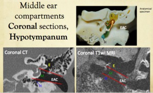
Fig. 8: Coronal sections (dry temporal bone, coronal CT, coronal T1wi MRI), Hypotympanum
References: Department of Radiology 1, Strasbourg University Hospital, Strasbourg University, France 2015
The Hypotympanum (H) is located below the level of the external auditory canal (EAC).
Coronal CT section,
T1W and HRT2W show the hypotympanum located below a line (red line) tangent to the floor of the EAC.
This red line goes below the internal ear
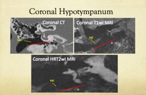
Fig. 9: Coronal sections (coronal CT, coronal T1wi MRI, coronal HRT2 wi MRI), Hypotympanum
References: Department of Radiology 1, Strasbourg University Hospital, Strasbourg University, France 2015
The middle ear is divided in three compartments on the axial section :
From anterior to posterior:
-Protympanum (P)
-Mesotympanum (M)
-Retrotympanum (R)
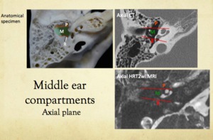
Fig. 10: Axial sections (dry temporal bone, axial CT, axial T2wi MRI)
References: Department of Radiology 1, Strasbourg University Hospital, Strasbourg University, France 2015
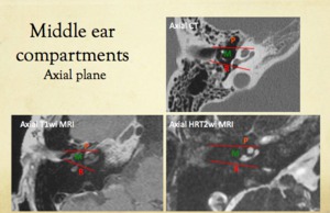
Fig. 11: Axial sections (axial CT, axial T1wi MRI, axial HRT2 wi MRI)
References: Department of Radiology 1, Strasbourg University Hospital, Strasbourg University, France 2015
The Protympanum (P,
orange) is anterior to a line tangent to theanterior wall of the external auditory canal (EAC).
The axial Protympanum (p) is the compartment located anteriorly to a line (red line) tangent to the anterior wall of the external auditory canal (EAC).Its upper part is located laterally to the geniculate ganglion fossa (gg).
Its lower part is located right before the tangent line to the anterior wall of the EAC and to the anterior part of the cochlea (co).
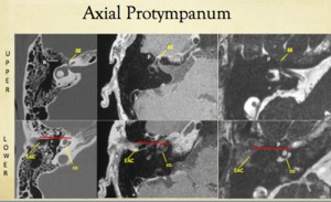
Fig. 12: Axial Protympanum (axial CT, axial T1 wi MRI, axial HRT2 wi MRI)
References: Department of Radiology 1, Strasbourg University Hospital, Strasbourg University, France 2015
The Mesotympanum (M,
green) is facing the tympanic membrane
laterally.
Medially,
the Mesotympanum is facing the basal turn of the
cochlea+ the inferior part of the vestibule.
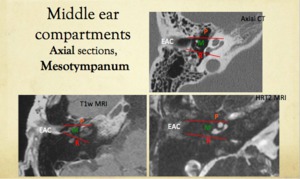
Fig. 13: Axial Mesotympanum (axial CT, axial T1 wi MRI, axial HRT2 wi MRI)
References: Department of Radiology 1, Strasbourg University Hospital, Strasbourg University, France 2015
The Retrotympanum (R,
red) is posterior to a line tangent to theposterior wall of the external auditory canal (EAC).
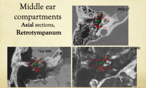
Fig. 14: Axial Retrotympanum (axial CT, axial T1 wi MRI, axial HRT2 MRI)
References: Department of Radiology 1, Strasbourg University Hospital, Strasbourg University, France 2015
The Retrotympanum (R) is located behind a tangent line to the posterior wall of the EAC laterally.
This line courses in front of the third (mastoid) portion of the facial nerve (CN7) and behind the basal turn of the cochlea (bt) + the round window (RW) medially.
It contains the facial recess (fr) laterally and the sinus tympani (st) medially to the mastoid portion of the facial nerve (CN7)
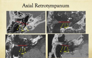
Fig. 15: Axial Retrotympanum (axial CT, axial HRT2 wi MEI, axial T1 wi MRI)
References: Department of Radiology 1, Strasbourg University Hospital, Strasbourg University, France 2015
SUMMARY :
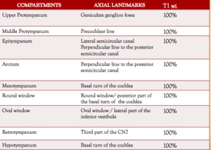
Fig. 16: Summary
References: Department of Radiology 1, Strasbourg University Hospital, Strasbourg University, France 2015
CLINICAL APPLICATIONS :
Case 1 =
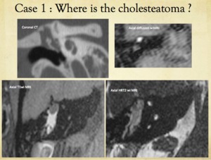
Fig. 17: Case 1 : where is the cholesteatoma ?
References: Department of Radiology 1, Strasbourg University Hospital, Strasbourg University, France 2015

Fig. 18: Case 1, CT explanation
References: Department of Radiology 1, Strasbourg University Hospital, Strasbourg University, France 2015
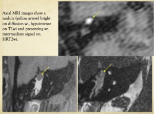
Fig. 19: Case 1 : MRI reading
References: Department of Radiology 1, Strasbourg University Hospital, Strasbourg University, France 2015
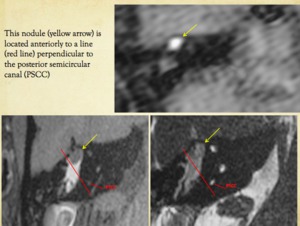
Fig. 20: Case 1 : MRI reading
References: Department of Radiology 1, Strasbourg University Hospital, Strasbourg University, France 2015
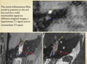
Fig. 21: Case 1 : MRI reading
References: Department of Radiology 1, Strasbourg University Hospital, Strasbourg University, France 2015
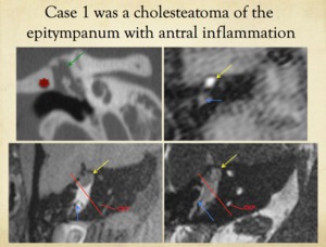
Fig. 22: Case 1 : correct localization of the lesion on MRI
References: Department of Radiology 1, Strasbourg University Hospital, Strasbourg University, France 2015
Case 2 =
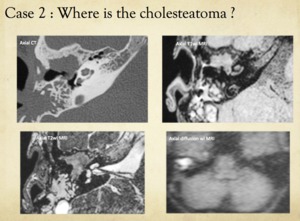
Fig. 23: Case 2 : where is the cholesteatoma ?
References: Department of Radiology 1, Strasbourg University Hospital, Strasbourg University, France 2015
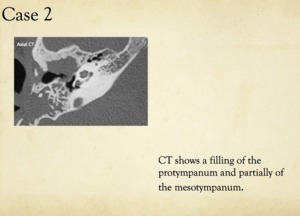
Fig. 24: Case 2 : CT reading
References: Department of Radiology 1, Strasbourg University Hospital, Strasbourg University, France 2015
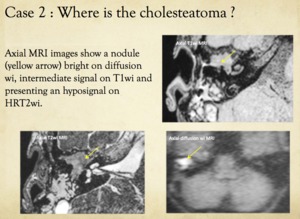
Fig. 25: Case 2 : MRI interpretation
References: Department of Radiology 1, Strasbourg University Hospital, Strasbourg University, France 2015
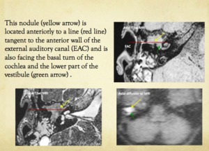
Fig. 26: Case 2 : MRI reading
References: Department of Radiology 1, Strasbourg University Hospital, Strasbourg University, France 2015
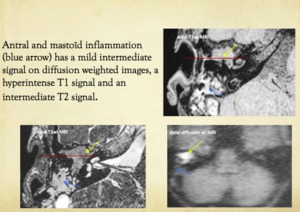
Fig. 27: Case 2 : MRI reading
References: Department of Radiology 1, Strasbourg University Hospital, Strasbourg University, France 2015
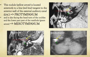
Fig. 28: Case 2 : MRI reading
References: Department of Radiology 1, Strasbourg University Hospital, Strasbourg University, France 2015
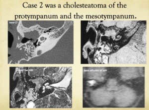
Fig. 29: Case 2 : localization of the lesion on MRI images
References: Department of Radiology 1, Strasbourg University Hospital, Strasbourg University, France 2015
