ECR 2016 / C-1843
Role of Imageology in hydrocephalus. Lets innovate to understand the not so beautiful ‘Sun setting’sign!
This poster is published under an open license. Please read the disclaimer for further details.
Congress:
ECR 2016
Poster Number:
C-1843
Type:
Educational Exhibit
Keywords:
CNS, Neuroradiology brain, CT, MR, MR-Functional imaging, Diagnostic procedure, Shunts, Localisation, Cerebrospinal fluid, Dilatation, Haemodynamics / Flow dynamics
Authors:
A. V. Nair1, R. Rajeshkannan2, S. Moorthy3, P. V. Ramachandran4; 1Trivandrum, kerala/IN, 2ERNAKULAM, Ke/IN, 3Kochi/IN, 4Cochin, Kerala/IN
DOI:
10.1594/ecr2016/C-1843

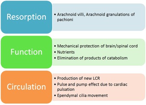
Fig. 1:
Properties of CSF

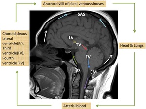
Fig. 2:
CSF circulation

Fig. 3:
Functions of CSF

Fig. 4:
Role of Imaging in hydrocephalus