Aims and objectives
To assess the variations of cerebral venous anatomy using non contrast MR venography.
To assess the potential pitfalls due to variations in diagnosis of cerebral venous pathological conditions
Methods and materials
Material & Method: This prospective observational study was carried out at “Apollo Hospitals”,
Bangalore,
from May 2014 & May 2015. Patients were selected as per inclusion and exclusion criteria.
Study area:Department of Radiodiagnosis,
Apollo Hospitals,
Bangalore.
Study population:All out-patient and in-patient undergoing MRI Brain study in the Department of Radio-Diagnosis of Apollo hospitals,
Bangalore.
Sample size:
Total number of patients- 100.
Data collection technique and tools:
Patients were selected as per inclusion and exclusion criteria. All patients (100) had undergone routine MR brain imaging sequences...
Results
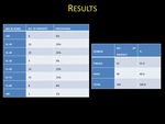
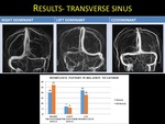
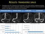
Transverse sinus asymmetry is the most common anatomical variation in cerebral venous system.
Hypoplasia ,
aplasia and flowgaps in transverse sinus are potential pitfalls in the diagnosis of transverse sinus thrombosis.
Sigmoid sinus calibre is in relation with the calibre of transverse sinus and internal jugular vein on same side .
Presence of sigmoid sinus variations with ipsilateral small internal jugular vein excludes sigmoid sinus thrombosis.
Though superior sagittal sinus is constant structure,
variations such as bifurcation,
Rostral hypogenesis are observed.
Flow gaps are usually...
Conclusion
Non contrast MR venography is one of the best and safe imaging modality to study the dural venous sinuses,
Vein of Galen,
internal cerebral vein ,
basal vein of Rosenthal and major anastomotic vein.
Variations in the calibre of the sinuses and flowgaps were more common in transverse sinuses ,
which are potential pitfalls for sinus thrombosis.
Sigmoid sinus asymmetry with ipsilateral small internal jugular vein excludes thrombosis of sigmoid sinus.
In superior sagittal sinus,
straight sinus and deep cerebral veins ,
flowgaps were less...
References
1. Lirng J.
Magnetic Resonance Venography of Intracranial Venous Diseases.
Journal of the Chinese Medical Association.
2010;73(6):289-291.
2. Krayenbuhl H.
Cerebral venous thrombosis: the diagnostic value of cerebral angiography.
Schweiz Arch Neurol Psychiatr.
1954; 74:261–287.
3. Ameri A,
Bousser MG.
Cerebral venous thrombosis.
Neurol Clin.
1992.
Feb;10(1):87-111.
4. Pillai LV,
Ambike DP,
Nirhale S,
Husainy SMK,
Pataskar S,
et al.
Cerebral venous thrombosis: An experience with anticoagulation with low molecular weight heparin.
Indian J Crit Care Med 2005; 9:14-18.
5. Ayanzen RH,
Bird CR,
Keller...


