
ECR 2018 / C-1324
Digital subtraction Neuroangiography: what a resident should know.
Congress:
ECR 2018
Poster Number:
C-1324
Type:
Educational Exhibit
Keywords:
Education and training, Structured reporting, Education, Arterial access, Conventional radiography, Catheter arteriography, Vascular, Neuroradiology brain, Interventional vascular
Authors:
S. Neelakantan1, R. Samant2, B. N. Reddy3, P. Reddy4, B. B. Das1, S. Viswamitra4; 1Bangalore, Karnataka/IN, 2Little Rock, Arkansas/US, 3Bangalore, ka/IN, 4Bangalore/IN
DOI:
10.1594/ecr2018/C-1324

Fig. 5:
Types of catheters used.
A: Angled vertebral catheter for routine cases
B:...

Fig. 6:
There is evidence of a blister like saccular ACOM aneurysm on left ICA...

Fig. 7:
Saccular ACOM aneurysm noted with narrow neck. Distal ACA filling is within...

Fig. 8:
Severe stenosis of carotid bifurcation and RICA origin are noted on the lateral...
 with diffuse nidus in the left cerebellar. Feeders from hemispheric branches of left Superior Cerebellar Artery, Posterior Inferior Cerebellar Artery (hemispheric and vermian branches). Intranidal pseudoaneurysm noted on the posterosuperior aspect of the nidus. Venous drainage is through multiple cerebellar veins and superior vermian vein into the straight sinus and transverse sinus References: Department of Radiology, SSSIHMS, Bangalore")
Fig. 9:
There is evidence of an arterio-venous malformation (SM score 3) with diffuse...
, distal right internal maxillary branches, right middle meningeal artery. Mass effect on the adjacent RICA - widening of C loop with mild luminal narrowing. Patchy tumor blush and cork-screw pattern is seen appearing in the early arterial phase, progressively increasing and persisting into the delayed venous phase. Hypoplastic A1 segment of the right ACA. References: Department of Radiology, SSSIHMS, Bangalore")
Fig. 10:
Axial T2 weighted MR image showing a right parasellar homogenously bright...

Fig. 11:
Patient with chronic headache and seizures for more than 10 years. There is...
 in the right frontal region fed by hypertrophied frontal branches of right ACA and MCA. Drainage is via a single cortical vein into the anterior third of superior sagittal sinus and also via cortical vein into spheno-parietal sinus and thence into right transverse-sigmoid junction. Post operative check angiogram shows no evidence of residual AVM / vessel spasm. References: Department of Radiology, SSSIHMS, Bangalore")
Fig. 12:
There is evidence of compact nidus AVM (SM score I) in the right frontal region...
. References: Department of Radiology, SSSIHMS, Bangalore")
Fig. 13:
There is evidence of a fuso-saccular aneurysm arising from the V4 segment of...

Fig. 14:
There is evidence of a dural vascular malformation with fistulous component at...

Fig. 15:
There is evidence of high flow direct carotico-cavernous fistula with rent...
 on left side being fed by Dural branches arising from cavernous segment right ICA, distal/ dural branches of right internal maxillary artery and middle meningeal. Drainage is antegrade; predominantly anterior through the superior ophthalmic vein into angular vein, into the facial and superficial temporal veins. References: Department of Radiology, SSSIHMS, Bangalore")
Fig. 16:
There is evidence of a slow flow right sided indirect CCF (Type D2) on left...
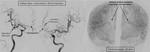
Fig. 17:
There is evidence of saccular DACA aneurysm arising at the bifurcation of ACA...
 at the apex. The superior cerebellar arteries and posterior cerebral arteries are noted separately from the sac. References: Department of Radiology, SSSIHMS, Bangalore")
Fig. 18:
Saccular basilar top aneurysm with eccentric outpouching (daughter aneurysm) at...

Fig. 19:
Susceptibility weighted MR image shows bilateral cerebellar parenchymal brush...
. Post endovascular and percutaneous embolization, check angiogram shows no tumour blush. References: Department of Radiology, SSSIHMS, Bangalore")
Fig. 20:
MR images shows a intensely enhancing soft tissue lesion widening the spheno-...

Table 1:
Anatomic structures of interest with specific angiographic views.