Many radiological signs are named by the things they resemble which may include inanimate objects,
food,
plants,
and even animals.
In this exhibition,
we will discuss and illustrate some of the most common radiological signs of a variety of radiological subspecialties and imaging modalities,
whose name was inspired by their resemblance with a diversity of animals.
Let’s get ready for a visual walk through the zoo!
Anteater nose sign
Calcaneonavicular coalition refers to a fusion between the calcaneus and navicular bones.
The anterior process of the calcaneus is elongated,
better visualized on lateral radiographs of the foot.
The elongated process resembles the long snout of an anteater.
[ Fig. 1 ] The finding may also be seen on oblique radiographs of the foot.
[10]
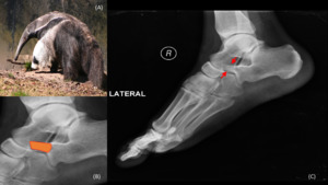
Fig. 1: (A) Giant Anteater at the Copenhagen Zoo, Denmark, on 2005. By Malene Thyssen (Own work) [GFDL (http://www.gnu.org/copyleft/fdl.html), CC-BY-SA-3.0 (http://creativecommons.org/licenses/by-sa/3.0/) or CC BY-SA 2.5 (https://creativecommons.org/licenses/by-sa/2.5)], via Wikimedia Commons. Lateral radiograph of the foot (B) with calcaneonavicular coalition. Lateral radiograph of the foot (C) with anterior tubular prolongation of the superior calcaneus which approaches or overlaps the navicular bone. This resembles the nose of an anteater.
Bat’s wings appearance
Perihilar opacifications that extend laterally are almost pathognomonic of airspace-filling processes.
These pattern of alveolar infiltrates are referred to as bat’s wing appearance or butterfly pattern of lung disease due to its resemblance to butterfly wings on chest x-ray.
[ Fig. 2 ] Airspace diseases that most commonly produce this pattern are pulmonary edema and pulmonary hemorrhage.
[4]
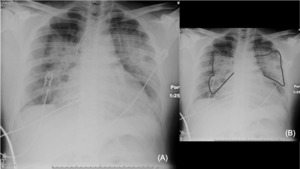
Fig. 2: Portable chest radiograph (A) with bilateral perihilar opacities. (B) Diagram over bilateral perihilar opacities showing the resemblance with bat’s wings (grey lines).
Bear paw sign
Xanthogranulomatous pyelonephritis is a chronic kidney infection,
usually with Proteus mirabilis,
and commonly involving an obstructed kidney.
The destroyed renal parenchyma is replaced by lipid-laden macrophages (xanthoma cells) and also fibrofatty replacement.
Patients present with flank pain and constitutional symptoms.
On abdominal CT and ultrasonography,
the affected kidney is atrophic and has hydronephrosis.
Either diffuse or localized,
it may mimic a renal mass.
On MR,
the renal calyces are dilated and abscesses replace the normal renal parenchyma.
The bear paw sign refers to the pattern created by the hypoattenuating renal calyces (replaced by fibro-fatty tissue) in a radial arrangement,
resembling the paw of a bear.
[4,11] [ Fig. 3 ]
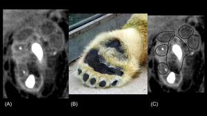
Fig. 3: Contrast-enhanced abdominopelvic CT scan, coronal views (A) and (C) on delayed phase with hypoattenuating renal calyces favoring xanthogranulomatous pyelonephritis (black lines). (B) Paw of a polar bear in the Karlsruhe zoo. By --Srvban 00:10, 28. Aug. 2009 (CEST) (Own work) [CC BY-SA 3.0 (https://creativecommons.org/licenses/by-sa/3.0), CC BY-SA 3.0 de (https://creativecommons.org/licenses/by-sa/3.0/de/deed.en) or GFDL (http://www.gnu.org/copyleft/fdl.html)], via Wikimedia Commons.
Bird’s beak sign
Achalasia is a motility disorder affecting the distal part of the esophagus.
In achalasia,
there is no peristalsis in the esophageal body and the lower esophageal sphincter is unable to relax due to marked increased resting pressure.
Secondary achalasia may be caused by Chagas disease and neoplasms obstructing the gastroesophageal junction.
Barium swallow illustrates the classic bird’s beak appearance of achalasia in which there is marked proximal esophageal dilatation,
and a stricture or tapered narrowing of the distal esophagus.
[ Fig. 4 ] The beaking pattern illustrated in achalasia has also been used to describe pyloric stenosis and colonic volvulus.
[4,11]
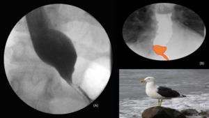
Fig. 4: (A) Barium swallow with tapering of the inferior esophagus in achalasia. (B) Diagram over barium swallow, showing the bird beak sign in achalasia. (C) Photograph of a black backed gull in Wellington Harbour, Wellington, New Zealand. I, Tony Wills [GFDL (http://www.gnu.org/copyleft/fdl.html), CC BY 2.5 (http://creativecommons.org/licenses/by/2.5) or CC BY-SA 3.0 (https://creativecommons.org/licenses/by-sa/3.0)], via Wikimedia Commons.
In sigmoid volvulus,
if a barium enema is performed it demonstrates the obstruction tapering to form a beak at the point where the twist occurs.
The beaking pattern seen on fluoroscopy is also referred to as bird of prey sign.
[4]
Butterfly glioma
Glioblastomas commonly affect both brain hemispheres,
specially affecting bilateral frontal lobes,
spreading through the corpus callosum.
On brain CT and MR images,
this bi-hemispheric involvement resembles the wings of a butterfly,
and hence glioblastomas are commonly referred to as butterfly gliomas.
[ Fig. 5 ] This butterfly appearance of disease has also been described for CNS lymphoma and tumefactive demyelinating lesions involving the corpus callosum.
[4,7]
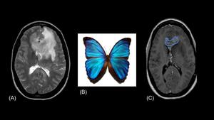
Fig. 5: Axial T2 (A) and T1 (C) views brain MRI showing a large region with abnormal signal intensity and patchy contrast enhancement involving the bilateral frontal lobes and the genu (blue line) and splenium of the corpus callosum (multifocal high-grade astrocytoma involving the bilateral cerebral hemispheres). (B) Photograph of a Blue Morpho butterfly. By Gregory Phillips - Own work, CC BY-SA 3.0, https://commons.wikimedia.org/w/index.php?curid=110687.
Butterfly vertebrae
Represents a sagittal cleft vertebrae,
this means that there is persistent notochord between the two non-fused lateral halves of the vertebral body.
The appearance on conventional radiographs and coronal CT images resembles the body of a butterfly,
with the non-fused halves representing the butterfly’s wings.
[ Fig. 6 ] Since this is a congenital defect,
the associated intervertebral discs develop concavities bilaterally to conform to the vertebral body.
This defect has been associated with spina bifida.
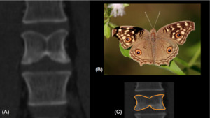
Fig. 6: Abdominopelvic CT scan, coronal view (A) on bone window, showing an incomplete anterior butterfly vertebrae at L4 level. (B) Junonia lemonias CC BY 2.5, https://commons.wikimedia.org/w/index.php?curid=469894. (C) Diagram over the L4 vertebrae delimiting the butterfly pattern (orange line).
Cobra head sign
Simple ureterocoele,
usually described incidentally on adults,
produces distal ureter (intravesicular) dilatation.
On contrast-enhanced studies,
this dilatation is surrounded by a lucent line (ureter wall) and such appearance is named the cobra head sign.
[ Fig. 7 ] Simple ureteroceles are caused by congenital prolapse of the distal ureter into the urinary bladder.
[4]
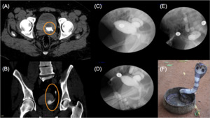
Fig. 7: Axial (A) and coronal (B) views of an abdominopelvic CT scan on delayed phase demonstrating a left ureterocele (orange circles). (C), (D) and (E)Cystourethrogram images with a contrast-filled outpouching consistent with a right everted ureterocele. Contrast reflux into the right ureter and pelvocaliceal system was evidenced showing significant ureteral tortuosity and hydroureteronephrosis. (F) Cobra head sign. By No machine-readable author provided. Jolle~commonswiki assumed (based on copyright claims). [GFDL (http://www.gnu.org/copyleft/fdl.html) or CC-BY-SA-3.0 (http://creativecommons.org/licenses/by-sa/3.0/)], via Wikimedia Commons.
Codfish vertebrae
Or just fish vertebrae,
refers to when there is bilateral compression of the vertebral body due to osteoporotic changes,
resulting in a biconcave vertebral body.
This biconcave appearance is termed codfish or fish vertebrae since the normal spine of a fish has that configuration.
[9]
Dromedary hump
May mimic a renal mass.
A dromedary hump represents a focal bulge on the renal cortex,
resembling the hump of a camel.
[ Fig. 8 ] It is a normal anatomical variant with normal renal function.
[4]
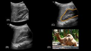
Fig. 8: Renal ultrasound (A) right kidney with normal size, shape and configuration. (B) and (C) are abdominal sonogram views in another patient with a right kidney dromedary hump (orange line). (D) Dromedary camel. By Rusty Clark from merritt usland FLA (Brevard Zoo at Viera FL) [CC BY 2.0 (http://creativecommons.org/licenses/by/2.0)], via Wikimedia Commons.
Feline esophagus
On barium swallow studies,
the normal esophageal mucosa is smooth.
When a patient presents with esophagitis or esophageal dysmotility disorders,
the multiple thick folds become evident due to the contraction of the longitudinal fibers in the muscularis mucosa.
[ Fig. 9 ] This appearance is normal in the esophagus of cats,
hence when described in humans it is referred to as feline esophagus.[4]

Fig. 9: Upper GI fluoroscopy demonstrating transient horizontal ridges throughout the esophagus, representing a feline esophagus, in a patient with history of gastroesophageal reflux disease.
Gooseneck sign
In endocardial cushion defects or ostium primum type of atrial septal defect,
the precursor of the interatrial and interventricular septa has an abnormal development.
There are different degrees of malformation,
from two separate atrioventricular valves,
to a single valve.
In left ventricular angiography,
the left ventricular outflow tract is elongated and resembles the appearance of a gooseneck.
Endocardial cushion defects are typically seen in patients with trisomy 21.
[4]
Gull-wing appearance
Erosive osteoarthritis is a subtype of osteoarthritis that usually affects female patients.
It involves the joints commonly affected by osteoarthritis,
but has the erosive and inflammatory components of inflammatory arthritis.
It has a predilection for the hands and causes joint space narrowing,
joint ankylosis and subchondral erosions.
On conventional radiographs of the hands,
the central subchondral erosions of the distal interphalangeal joints in combination with loss of joint space and marginal osteophyte formation results in a lucency that resembles the silhouette of a sea gull,
hence the name gull wing appearance.
[ Fig. 10 ] Erosive osteoarthritis has a good prognosis and treatment is usually conservative.
[11]
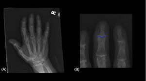
Fig. 10: Hand AP view (A) in a patient with erosive osteoarthritis with gullwing appearance of the distal interphalangeal (DIP) joints due to central subchondral sclerosis and marginal osteophyte formation. (B) Diagram over the third DIP representing the gullwing appearance (blue line).
Lobster claw sign
Papillary necrosis is ischemic necrosis of the tips of the medullary pyramids and loss of the renal parenchymal tissue.
Patients present with gross hematuria and renal insufficiency.
Causes of papillary necrosis include diabetes mellitus,
NSAID use,
sickle cell anemia and infection,
among others.
On CT urography,
there is contrast pooling / collection in the papilla.
One of the common imaging findings is contrast enhancing only the peripheral zone of the papilla creating an appearance of the claw of a lobster.
[11] [ Fig. 11 ]
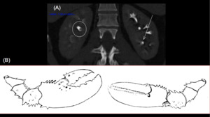
Fig. 11: Abdominopelvic CT scan with IV contrast coronal view (A) with forniceal excavation of a renal papilla seen in the interpolar region of the right kidney (circle), in a patient with renal papillary necrosis. Additionally, there is a central focal excavation of a renal papilla seen in the inferior pole of the left kidney (white arrow). (B) Lobster claws. Unknown [Public domain], via Wikimedia Commons.
Panda sign
When there is inflammation of the parotid glands,
nasopharynx and lacrimal glands,
on Gallium scans the affected areas look dark,
resembling the markings on the face of a panda.
Although not specific,
it almost always describes on patients with sarcoidosis.
[11]
Scottie dog sign
Spondylolysis is an interruption or defect in the pars interarticularis portion of the posterior elements (lamina) of the vertebra.
On conventional oblique radiographs or oblique CT scans of the lumbar spine,
the normal,
un-interrupted,
pars interarticularis resembles the body of a Scottie dog.
The inferior articulating facet represents the dog’s front leg,
the contralateral inferior articulating facet represents the rear leg; the lamina forms the body; the pedicle the eye; the superior articulating facet is the ear,
the contralateral superior articulating facet is the dog’s tail; and finally,
the transverse process forms the dog’s nose.
[ Fig. 12 ] When there is a pars defect,
a collar forms around the Scottie dog’s neck.
[4,
10]
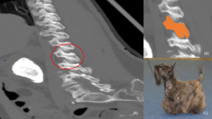
Fig. 12: Oblique CT scan (A) of the lumbar spine. The posterior elements of the vertebra form the figure of a Scotty dog (red circle). (B) Scotty dog diagram over oblique CT scan of the lumbar spine. Nose is formed by the transverse process; pedicle forms the eye; interior articular facet is the front leg; superior articular facet represents the ear; and the pars interarticularis is equivalent to the neck of the dog. (C) Brindle Scottish Terrier. By Томасина (Own work) [CC BY-SA 3.0 (https://creativecommons.org/licenses/by-sa/3.0)], via Wikimedia Commons.
Staghorn calculi
Urinary calculi vary in size and shape,
and approximately 85% are visualized on conventional radiographs.
On oblique radiographs,
their posterior position differentiates renal stones from gallstones.
A large calculus that occupies the renal collecting system is referred to as a staghorn calculus due to its shape resembling the horns of a stag and also the coral with the same name.
[ Fig. 13 ,
Fig. 14 ] Staghorn calculi are usually composed of struvite.
Commonly associated with recurrent urinary tract infections and present with flank pain,
fever,
hematuria and even abscess formation.
Surgical treatment is warranted to prevent progression to xanthogranulomatous pyelonephritis.
[4]
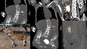
Fig. 13: Coronal view of an abdominopelvic CT scan (A) with a large radiopaque calculi within the left kidney with cortical thinning, conforming to the renal pelvis and calyces. (B) Photograph of a white-tailed deer. By USDA photo by Scott Bauer [Public domain], via Wikimedia Commons. (C) Diagram over the left kidney staghorn calculi. Coronal view of an abdominopelvic CT scan (D) with a right staghorn calculi. Coronal view of an abdominopelvic CT scan (E) with a right staghorn calculi, on bone window.
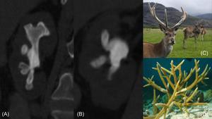
Fig. 14: Abdominopelvic CT scan, coronal views (A) and (B) showing a right kidney staghorn calculus. (C) Red deer stag. By Mehmet Karatay (Own work) [CC BY-SA 3.0 (https://creativecommons.org/licenses/by-sa/3.0) or GFDL (http://www.gnu.org/copyleft/fdl.html)], via Wikimedia Commons. (D) Staghorn coral. Adona9 at the English language Wikipedia [GFDL (http://www.gnu.org/copyleft/fdl.html) or CC-BY-SA-3.0 (http://creativecommons.org/licenses/by-sa/3.0/)], via Wikimedia Commons.
Swan neck deformity
Rheumatoid arthritis is a symmetric,
erosive inflammatory polyarticular arthropathy causing soft tissue swelling and uniform cartilage destruction.
Most common involved joints are: wrist joint,
proximal interphalangeal and metacarpophalangeal joints,
shoulder,
knee,
hips,
metatarsophalangeal joints,
and upper cervical spine.
It is considered a multisystemic disease,
but joint involvement is the predominant element.
When involving the hands,
there may be hyperextension of the proximal interphalangeal joint with compensatory hyperflexion of the distal interphalangeal joint.
This deformity is called a swan-neck deformity due to its similarity with the neck of a swan.
It may also be seen in scleroderma,
psoriatic arthritis and systemic lupus erythematosus.
[10]
Talar beak
Tarsal coalition occurs when there is a fusion between bones of the hindfoot or midfoot.
Talocalcaneal (subtalar) and calcaneonavicular are the two most common tarsal coalitions.
When a large osteophyte forms at the distal talus causing a superior projection of the distal aspect of the talus,
it is known as the talar beak sign,
as its imaging characteristics resemble the beak of a bird.
It is form by the excess motion / abnormal stress of the talonavicular joint.
Talar beaks are more common on subtalar coalitions than calcaneonavicular coalitions.
[10]

 Giant Anteater at the Copenhagen Zoo, Denmark, on 2005. By Malene Thyssen (Own work) [GFDL (http://www.gnu.org/copyleft/fdl.html), CC-BY-SA-3.0 (http://creativecommons.org/licenses/by-sa/3.0/) or CC BY-SA 2.5 (https://creativecommons.org/licenses/by-sa/2.5)], via Wikimedia Commons. Lateral radiograph of the foot (B) with calcaneonavicular coalition. Lateral radiograph of the foot (C) with anterior tubular prolongation of the superior calcaneus which approaches or overlaps the navicular bone. This resembles the nose of an anteater.")
 with bilateral perihilar opacities. (B) Diagram over bilateral perihilar opacities showing the resemblance with bat’s wings (grey lines).")
 and (C) on delayed phase with hypoattenuating renal calyces favoring xanthogranulomatous pyelonephritis (black lines). (B) Paw of a polar bear in the Karlsruhe zoo. By --Srvban 00:10, 28. Aug. 2009 (CEST) (Own work) [CC BY-SA 3.0 (https://creativecommons.org/licenses/by-sa/3.0), CC BY-SA 3.0 de (https://creativecommons.org/licenses/by-sa/3.0/de/deed.en) or GFDL (http://www.gnu.org/copyleft/fdl.html)], via Wikimedia Commons.")
 Barium swallow with tapering of the inferior esophagus in achalasia. (B) Diagram over barium swallow, showing the bird beak sign in achalasia. (C) Photograph of a black backed gull in Wellington Harbour, Wellington, New Zealand. I, Tony Wills [GFDL (http://www.gnu.org/copyleft/fdl.html), CC BY 2.5 (http://creativecommons.org/licenses/by/2.5) or CC BY-SA 3.0 (https://creativecommons.org/licenses/by-sa/3.0)], via Wikimedia Commons.")
 and T1 (C) views brain MRI showing a large region with abnormal signal intensity and patchy contrast enhancement involving the bilateral frontal lobes and the genu (blue line) and splenium of the corpus callosum (multifocal high-grade astrocytoma involving the bilateral cerebral hemispheres). (B) Photograph of a Blue Morpho butterfly. By Gregory Phillips - Own work, CC BY-SA 3.0, https://commons.wikimedia.org/w/index.php?curid=110687.")
 on bone window, showing an incomplete anterior butterfly vertebrae at L4 level. (B) Junonia lemonias CC BY 2.5, https://commons.wikimedia.org/w/index.php?curid=469894. (C) Diagram over the L4 vertebrae delimiting the butterfly pattern (orange line).")
 and coronal (B) views of an abdominopelvic CT scan on delayed phase demonstrating a left ureterocele (orange circles). (C), (D) and (E)Cystourethrogram images with a contrast-filled outpouching consistent with a right everted ureterocele. Contrast reflux into the right ureter and pelvocaliceal system was evidenced showing significant ureteral tortuosity and hydroureteronephrosis. (F) Cobra head sign. By No machine-readable author provided. Jolle~commonswiki assumed (based on copyright claims). [GFDL (http://www.gnu.org/copyleft/fdl.html) or CC-BY-SA-3.0 (http://creativecommons.org/licenses/by-sa/3.0/)], via Wikimedia Commons.")
 right kidney with normal size, shape and configuration. (B) and (C) are abdominal sonogram views in another patient with a right kidney dromedary hump (orange line). (D) Dromedary camel. By Rusty Clark from merritt usland FLA (Brevard Zoo at Viera FL) [CC BY 2.0 (http://creativecommons.org/licenses/by/2.0)], via Wikimedia Commons.")
 in a patient with erosive osteoarthritis with gullwing appearance of the distal interphalangeal (DIP) joints due to central subchondral sclerosis and marginal osteophyte formation. (B) Diagram over the third DIP representing the gullwing appearance (blue line).")
 with forniceal excavation of a renal papilla seen in the interpolar region of the right kidney (circle), in a patient with renal papillary necrosis. Additionally, there is a central focal excavation of a renal papilla seen in the inferior pole of the left kidney (white arrow). (B) Lobster claws. Unknown [Public domain], via Wikimedia Commons.")
 of the lumbar spine. The posterior elements of the vertebra form the figure of a Scotty dog (red circle). (B) Scotty dog diagram over oblique CT scan of the lumbar spine. Nose is formed by the transverse process; pedicle forms the eye; interior articular facet is the front leg; superior articular facet represents the ear; and the pars interarticularis is equivalent to the neck of the dog. (C) Brindle Scottish Terrier. By Томасина (Own work) [CC BY-SA 3.0 (https://creativecommons.org/licenses/by-sa/3.0)], via Wikimedia Commons.")
 with a large radiopaque calculi within the left kidney with cortical thinning, conforming to the renal pelvis and calyces. (B) Photograph of a white-tailed deer. By USDA photo by Scott Bauer [Public domain], via Wikimedia Commons. (C) Diagram over the left kidney staghorn calculi. Coronal view of an abdominopelvic CT scan (D) with a right staghorn calculi. Coronal view of an abdominopelvic CT scan (E) with a right staghorn calculi, on bone window.")
 and (B) showing a right kidney staghorn calculus. (C) Red deer stag. By Mehmet Karatay (Own work) [CC BY-SA 3.0 (https://creativecommons.org/licenses/by-sa/3.0) or GFDL (http://www.gnu.org/copyleft/fdl.html)], via Wikimedia Commons. (D) Staghorn coral. Adona9 at the English language Wikipedia [GFDL (http://www.gnu.org/copyleft/fdl.html) or CC-BY-SA-3.0 (http://creativecommons.org/licenses/by-sa/3.0/)], via Wikimedia Commons.")
