ECR 2018 / C-1742
CT of hepatocellular carcinoma in non-alcoholic fatty liver disease: imaging characteristics and inter-rater agreement
Congress:
ECR 2018
Poster Number:
C-1742
Type:
Scientific Exhibit
Keywords:
Liver, Abdomen, Oncology, CT, Diagnostic procedure, Observer performance, Cancer, Neoplasia
Authors:
I. Garg, S. Thompson, E. Ehman, S. P. Sheedy, A. Khandelwal, C. A. Bookwalter, T. Mounajjed, S. K. Venkatesh; Rochester, Mn/US
DOI:
10.1594/ecr2018/C-1742

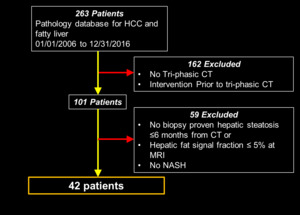
Fig. 1:
Flow chart showing selection of patients with HCC in NAFLD
 shows APHE, no PVWO but with DPWO and no capsule. Histology showed a well differentiated HCC.")
Fig. 2:
HCC in NAFLD.
70-year-old male with BMI of 30.7, diabetes and dyslipidemia....
 showing peripheral APHE, no PVWO and some DPWO. Note absence of capsule and a central region of hypo enhancement suggestive of necrosis (*). Resected Specimen showing HCC with central necrosis (dashed arrow. Histology showed a well-differentiated HCC.")
Fig. 3:
HCC in NAFLD.
75-year-old male with HCC in non-cirrhotic liver with mild...
 shows heterogeneous APHE with no PVWO, DPWO or capsule.")
Fig. 4:
HCC in NAFLD.
73-year-old male patient with HCC in severe steatohepatitis...

Fig. 5:
HCC in NAFLD (Inter reader agreement.
47-year-old female with HCC in...
 shows APHE, no PVWO but some DPWO and no capsule. Histology confirmed a well differentiated HCC.")
Fig. 6:
HCC in NAFLD
80-year-old male with HCC in non-cirrhotic NAFLD with mild...