Aims and objectives
Ultrasound is an inexpensive,
non-invasive and widely used imaging modality which has been demonstrated to be a valuable tool in the detection and monitoring of breast lesions and cancer,
especially when it is combined with screening mammography in women with dense breasts(1).
However,
the power of breast ultrasound can be limited by its inherent technical difficulties and inter-operator variability.
One study revealed that less than half of the lesions measuring 5 mm or smaller were identified by all experienced examiners(2).
The landmark study ACRIN 6666...
Methods and materials
Institutional review board approval from the North Shore University Health System/Evanston,
IL,
USA,
was obtained and all participants signed a written consent before the data collection.
Ultrasound examinations were performed with a GE Logic-E9 ultrasound machine (General Electric,
Milwaukee,
WI USA) integrated with the automated breast ultrasound system BVN Model G-1000.
Six women aged between 26 and 57 years old and with known benign or probably benign masses measuring 0.5-2 cm in maximum dimensions were scanned by two radiologists with at least five years of...
Results
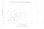
The 3D and 2D MDs positively correlated with PCD,
PAD and BRD,
independent from the other probe-position associated variables (ρ = 0.49,
0.23,
p<0.0001,
p<0.0001; ρ = 0.30,
0.40,
p=0.0002,
p<0.0001; and ρ = 0.28,
0.23,
p=0.0004,
p=0.0005 respectively).
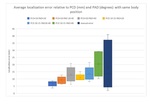
The 3D average MD with the probe and body positions differences in close range,
PCD <10 mm,
BRD <10 degrees and PAD <20 degrees was 7.9 mm (standard deviation,
sd=3.5mm,
n=27).
The smallest average 3D localization error (7.6mm) was obtained with the scan planes parallel between...
Conclusion
Our findings show that the displacement of small masses,
relative to nipple and body planes correlates with the changes in probe position and orientation,
as well as changes in body position on the exam table.
Large target displacements of a few cm were associated with larger differences in the probe and body positions between exams which are probably responsible for the limited reproducibility of lesions localization with the current manual annotations.
A localization error of approximately 7 mm can be obtained by matching the body...
References
1.
Kelly,
K.,
Dean,
J.,
Comulada,
W.
& Lee,
S.J.
Breast cancer detection using automated whole breast ultrasound and mammography in radiographically dense breasts.
European Radiology 20,
734-742 (2010).
2.
Mandelson,
M.T.
et al.
Breast Density as a Predictor of Mammographic Detection: Comparison of Interval- and Screen-Detected Cancers.
Journal of the National Cancer Institute 92,
1081-1087 (2000).
3.
Barr,
R.G.,
Zhang,
Z.,
Cormack,
J.B.,
Mendelson,
E.B.
& Berg,
W.A.; Probably Benign Lesions at Screening Breast US in a Population with Elevated Risk: Prevalence and Rate...


