ECR 2018 / C-2196

Echinococcosis from head to toe: A Pictorial review
Congress:
ECR 2018
Poster Number:
C-2196
Type:
Educational Exhibit
Keywords:
Abdomen, Lung, CT, MR, Diagnostic procedure, Infection
Authors:
S. Patwari, R. V. Helavar, R. Govindappa, M. KUMAR, H. C. Chadaga; Bangalore/IN
DOI:
10.1594/ecr2018/C-2196

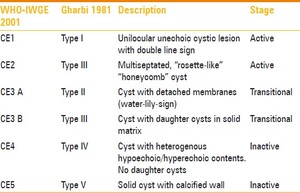
Fig. 3:
CLASSIFICATION OF HYDATID CYSTS

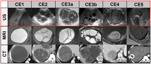
Fig. 4:
CLASSIFICATION OF HYDATID CYSTS
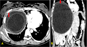
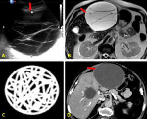
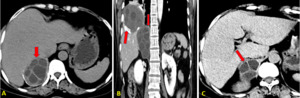
 Coronal CT scan demonstrates trilaminar pattern of the hepatic hydatid cyst with separation of the endocyst and fine peripheral enhancement of the cystic walls.")
Fig. 5:
Hepatic Hydatid cyst.
Axial CT scan (b) Coronal CT scan demonstrates...
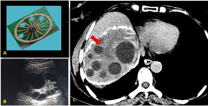
 USG image, (B) axial T2 MRI (C) representative pic (D) Axial CT demonstrating cyst with a predominantly thick membranes.")
Fig. 6:
(A) USG image, (B) axial T2 MRI (C) representative pic (D) Axial CT...

Fig. 7:
Hepatic hydatid with cart wheel like appearance.
Axial CT scan of liver and...
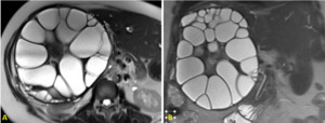
 Axial and (B) Coronal T2w MRI of liver demonstrates hepatic hydatid cyst with multiple daughter cysts in right lobe of liver.")
Fig. 8:
A) Axial and (B) Coronal T2w MRI of liver demonstrates hepatic hydatid cyst...
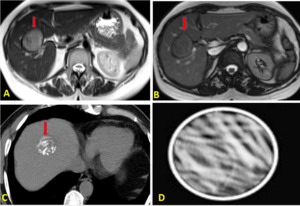
 and (B) Axial MRI, (C) Axial CT (D) representative image demonstrating cyst with calcified wall and internal contents, representing an involuting/ degenerating cyst.")
Fig. 9:
(A) and (B) Axial MRI, (C) Axial CT (D) representative image demonstrating cyst...
 Axial CT scans (D) Coronal CT scan images demonstrates thick walled hydatid cyst in the segment II and III of the liver with multiple enhancing internal septations seen communicating with the left hepatic duct. Similar lesions in the segment VIII . Multiple intraductal hydatid cysts causing upstream biliary radical dilatation.")
Fig. 10:
Biliary hydatid disease.
(A,B,C) Axial CT scans (D) Coronal CT scan images...

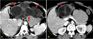
Fig. 11:
Adrenal hydatic cyst.
Axial and coronal, pre and post contrast CT scan...

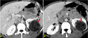
Fig. 12:
Renal hydatid. Axial CT scan with thick walled cyst with internal membranes...

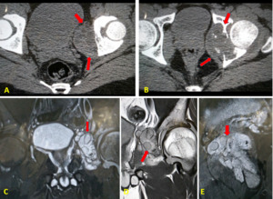
Fig. 13:
Intraperitoneal hydatid cysts
Axial CT scans demonstrates partial destruction of the acetabulum with adjacent soft tissue component with cystic areas within.
(C,D,E) MRI images in same patient demonstrates Cystic lesion with daughter cysts within suggestive of bony hydatid cyst.")
Fig. 14:
BONE HYDATID CYST.
(A,B)Axial CT scans demonstrates partial destruction of...
 Representative image.")
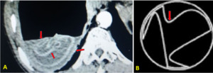
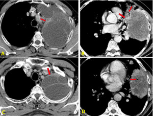
Fig. 15:
LUNG HYDATID CYST.
Axial CT showing pulmonary hydatid with floating internal...
, Image D showing air within cyst secondary to bronchial communication")
Fig. 16:
Pulmonary hydatid with intrabronchial communication (A,B,C), Image D showing...

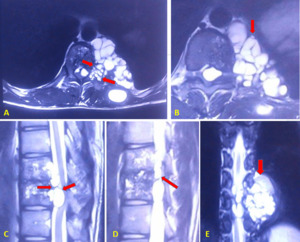
Fig. 17:
SPINAL HYDATID CYST

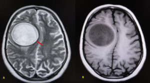
Fig. 18:
BRAIN HYDATID CYST.