ECR 2018 / C-2238
Commonest locations of challenging TB bug in spine
Congress:
ECR 2018
Poster Number:
C-2238
Type:
Educational Exhibit
Keywords:
Infection, Education, MR, Musculoskeletal system, Musculoskeletal spine
Authors:
S. Arooj1, M. farooq1, S. Kadri2, K. Qadir3; 1Karachi/PK, 2Karachi, karachi/PK, 3Karachi, Si/PK
DOI:
10.1594/ecr2018/C-2238

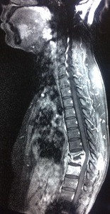
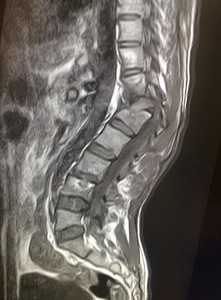
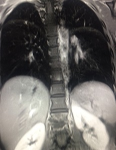
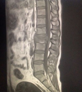
Fig. 1:
57 years old male with backache. Intraosseous tb abscesses are seen in lower...

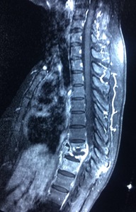
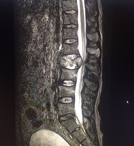
Fig. 2:
57 years old male with backache. Re demonstration of intra osseous TB abscesses...

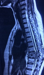
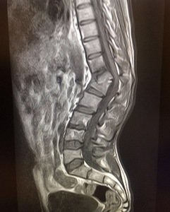
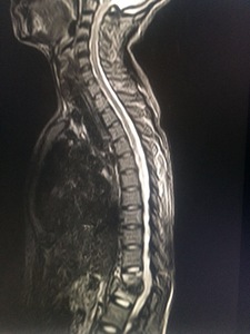
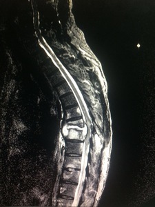
Fig. 3:
Same patient. Lower dorsal vertebral bodies show compression/ collapse with...

Fig. 4:
Same patient, similar T2WI findings as mentioned in fig.3

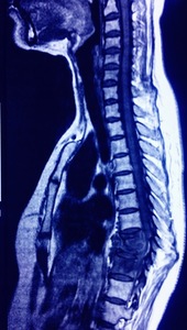
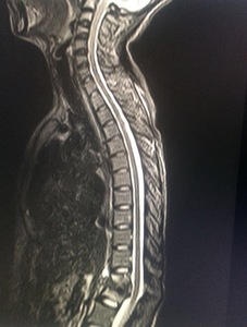
Fig. 5:
Same patient, Marrow replacement lower dorsal vertebrae, hypointense on T1WI....
")
Fig. 6:
Case 2.
23 years old male with history of fall two years back. complains of...

Fig. 7:
Case 2.
23 years old male with history of fall two years back. complains of...

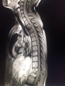
Fig. 8:
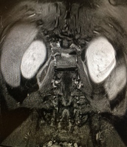
Same patient, post contrast coronal T1WI show oblique right paravertebral...

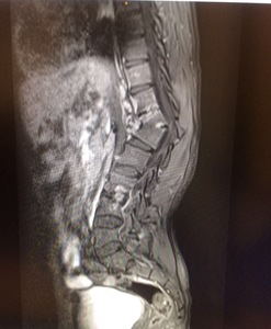
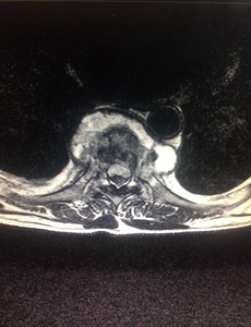
Fig. 9:
Same patient, post contrast T1WI show enhancement of endplates with...

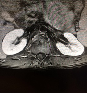
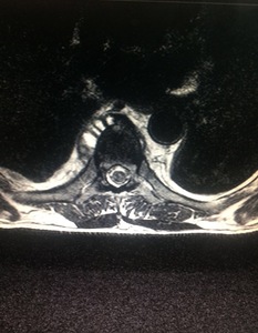
Fig. 10:
Same patient, post contrast axial T1WI show right paravertebral components with...

Fig. 11:
26 year old female with bilateral lower limb weakness. T2WI show compression/...

Fig. 12:
26 year old female with bilateral lower limb weakness. T2WI show compression/...

Fig. 13:
26 year old female with bilateral lower limb weakness. Post contrast sagittal...

Fig. 14:
26 year old female with bilateral lower limb weakness. Coonal T1WI show...

Fig. 15:
T1W hypointense marrow signals with involvement of multiple vertebral bodies in...

Fig. 16:
T2W axial images show bilateral paravertebral abscesses, hyperintense on T2W...

Fig. 17:
T2W hyperintense paravertebral components in a 15 year old female

Fig. 18:
T2W hyperintense marrow signals with involvement of multiple vertebral bodies...

Fig. 19:
40 years old labourer with low grade fever and backache.Hypointense L2...

Fig. 20:
40 years old labourer with low grade fever and backache.Hyperintense L2...