Type:
Educational Exhibit
Keywords:
Congenital, Chronic obstructive airways disease, Technical aspects, Normal variants, CT, Conventional radiography, Thorax
Authors:
J. D. Oliveira1, I. Martins2, A. I. Coutinho Santos2; 1Amadora/PT, 2Lisboa/PT
DOI:
10.1594/ecr2018/C-2348
Background
On chest radiography,
unilateral hyperlucency is described as a darker appearance of one side of the thorax,
due to increased x-ray transmission.
It is an uncommon finding that could not have clinical relevance,
being the result of technical factors or wrong positioning,
but could also be the radiological expression of various pathology,
either congenital or acquired,
ranging from benign to potentially life-threatening diseases.
The first step in the evaluation is to check on radiologic technique and positioning,
since the most common causes of a unilateral hyperlucent hemithorax do not reflect an intrinsic abnormality itself.
When faulty radiologic technique or poor patient positioning are excluded,
it may be useful to consider the potential causes by the structures they involve,
and thus as the result from asymmetric absence of chest soft tissues,
changes located to the pleura or the pleural space,
changes related to the pulmonary parenchyma/ventilation or by changes in the pulmonary vascularity/perfusion.
Since vascular structures are mainly responsible for the radiologic texture of the normal lung,
any change in the pulmonary vascularity results in loss of the lung opacity.
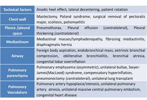
Fig. 1: Differential diagnosis of unilateral hypertranslucent hemithorax.
