Now that we have reviewed the key concepts about the anatomy and radiologic anatomy of the mesentery,
we can approach the mesentery pathologies by dividing them into primary and secondary.

Table 1: Table of contents
Primary
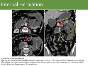
Fig. 11: Left internal paraduodenal hernia
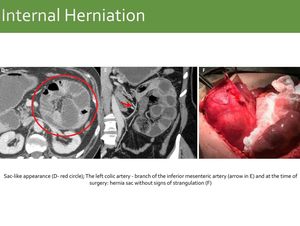
Fig. 12: Same case as Fig. 11: Left internal paraduodenal hernia
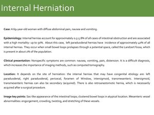
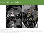
Fig. 13: Internal Herniation
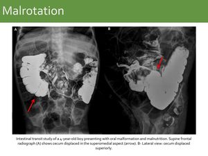
Fig. 14: Malrotation: Case 1.
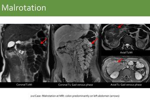
Fig. 15: Malrotation Case 2.
References: Fleury Medicina Diagnóstica, São Paulo, Brazil

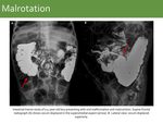
Fig. 16: Malrotation
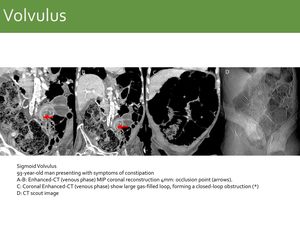
Fig. 17: Sigmoid Volvulus
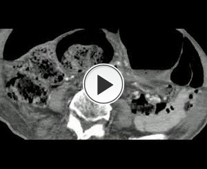
Fig. 18: Same case as Fig. 17: Sigmoid Volvulus Video - specific CT sign for volvulus is the whirl sign: vessels twisted like a whirlwind in the center of the bowel twist.

Fig. 19: Volvulus
- Vascular - Mesenteric bleeding/hematoma
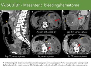
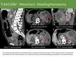
Fig. 20: Vascular - Mesenteric bleeding/hematoma

Fig. 21: Vascular - Mesenteric bleeding/hematoma
- Vascular - Mesenteric ischemia
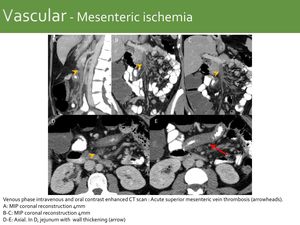
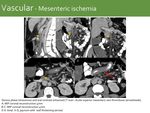
Fig. 22: Vascular - Mesenteric ischemia

Fig. 23: Vascular - Mesenteric ischemia
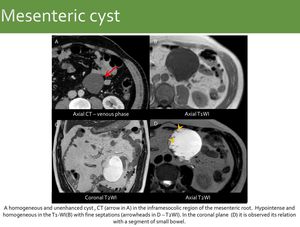
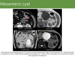
Fig. 24: Mesenteric cyst
References: Fleury Medicina Diagnóstica, São Paulo, Brazil
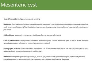
Fig. 25: Mesenteric cyst
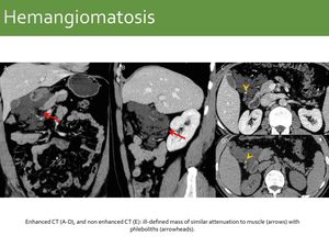
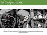
Fig. 26: Hemangiomatosis
References: Diagnósticos da América S.A. DASA. São Paulo, SP, Brazil
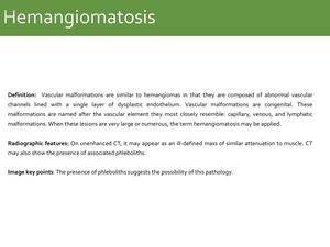
Fig. 27: Hemangiomatosis
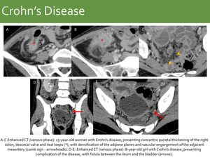
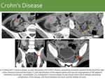
Fig. 28: Crohn’s Disease

Fig. 29: Crohn’s Disease
Primary Neoplasms
Tumors with primary manifestation in the peritoneum in the absence of a visceral site of origin.
They arise from mesothelial cells,
submesothelial mesenchymal cells,
and uncommitted stem cells .
Because the origins of some primary peritoneal tumors are obscure,
these lesions are difficult to classify precisely.
Mesothelial tumors: peritoneal malignant mesothelioma ,
well-differentiated papillary mesothelioma, multicystic mesothelioma and adenomatoid tumor.
Epithelial tumors: primary peritoneal serous carcinoma and primary peritoneal serous borderline tumor.
Smooth muscle tumor: leiomyomatosis peritonealis disseminata.
Tumors of uncertain origin: desmoplastic small round cell tumor and solitary fibrous tumor.
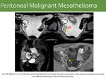
- Peritoneal Malignant Mesothelioma
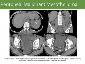
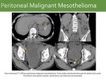
Fig. 30: Mesothelioma
References: Fleury Medicina Diagnóstica, São Paulo, Brazil
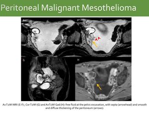
Fig. 31: Same case as Fig. 30: Mesothelioma
References: Fleury Medicina Diagnóstica, São Paulo, Brazil
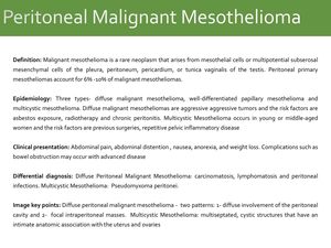
Fig. 32: Peritoneal Malignant Mesothelioma
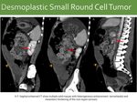
- Desmoplastic Small Round Cell Tumor
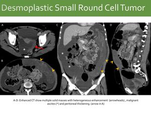
Fig. 33: Desmoplastic Small Round Cell Tumor
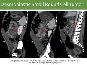
Fig. 34: Same case as Fig. 33: Desmoplastic Small Round Cell Tumor
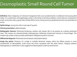
Fig. 35: Desmoplastic Small Round Cell Tumor
Primary- Inflammatory
- Sclerosing mesenteritis - mesenteric panniculitis
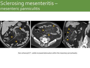
Fig. 36: Sclerosing mesenteritis – mesenteric panniculitis
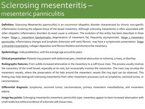
Fig. 37: Sclerosing mesenteritis – mesenteric panniculitis
- Sclerosing mesenteritis – retractile mesenteritis IgG4 related
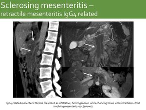
Fig. 38: Sclerosing mesenteritis – Retractile mesenteritis IgG4 related

Fig. 39: Sclerosing mesenteritis – retractile mesenteritis IgG4 related
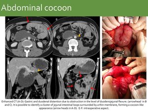
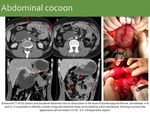
Fig. 40: Abdominal cocoon
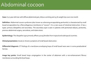
Fig. 41: Abdominal cocoon
Secondary Neoplasms
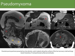
Fig. 42: Pseudomyxoma

Fig. 43: Pseudomyxoma
- Lymphomatosis x Carcinomatosis x Sarcomatosis
Conditions with overlapping characteristics:
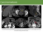
Carcinomatosis: Peritoneal and omental seeding are known sites of dissemination of metastatic carcinoma,
most commonly arising from the ovary,
colon and stomach.
Lymphadenopathy is usually located around the primary tumor.
Ascites is usually marked.
Lymphomatosis: omental caking with homogeneous bulky masses,
in addition to a diffuse distribution of enlarged lymph nodes.
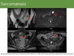
Sarcomatosis: may present bulky masses,
but they are frequently heterogeneous,
hypervascular and may be associated with hemoperitoneum.
Lymphnode enlargement is rare.
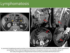
Fig. 44: Lymphomatosis

Fig. 45: Lymphomatosis
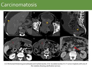
Fig. 46: Carcinomatosis

Fig. 47: Carcinomatosis
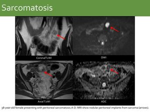
Fig. 48: Sarcomatosis
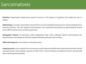
Fig. 49: Sarcomatosis
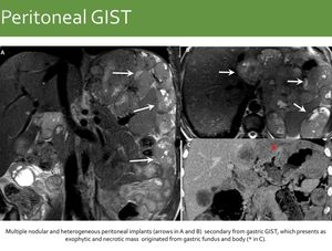
Fig. 50: Peritoneal GIST

Fig. 51: Peritoneal GIST
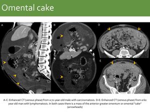
Fig. 52: Omental cake

Fig. 53: Omental cake
Secondary - Infectious
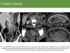
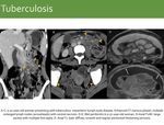
Fig. 54: Tuberculosis

Fig. 55: Tuberculosis
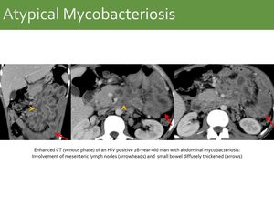
Fig. 56: Atypical Mycobacteriosis

Fig. 57: Atypical Mycobacteriosis
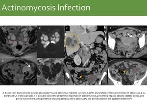
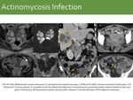
Fig. 58: Actinomycosis Infection
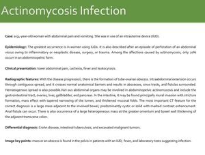
Fig. 59: Actinomycosis Infection
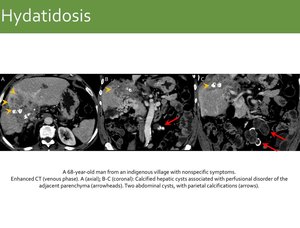
Fig. 60: Hydatidosis

Fig. 61: Hydatidosis
Secondary - Miscellany
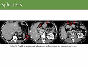
Fig. 62: Splenosis

Fig. 63: Splenosis


