ECR 2018 / C-2816

The Mesenteric Organ? - New concepts and a novel radiological perspective on its disease
Congress:
ECR 2018
Poster Number:
C-2816
Type:
Educational Exhibit
Keywords:
Abdomen, MR, CT, Education, Education and training, Mesentery, Peritoneum
Authors:
H. R. F. Dalla Pria1, F. Velloni1, R. A. Santiago1, M. S. Zacarias1, L. F. D. Silva1, F. Tamamoto1, A. C. Von Atzingen2, U. S. Torres1, G. D'Ippolito1; 1São Paulo/BR, 2Pouso Alegre/BR
DOI:
10.1594/ecr2018/C-2816

Fig. 1:
The new mesentery
. The small bowel mesentery has a "mesenteric root" at the origin of the superior mesenteric artery (SMA): the small bowel mesentery then fans out from the root region, where the SMA (red arrow) suspends it in the posterior abdominal wall to terminal ileum.")
Fig. 2:
Mesenteric root (orange). The small bowel mesentery has a "mesenteric root" at...
 with the superior mesenteric branches (vascular markings")
Fig. 3:
Small bowel mesentery (purple) with the superior mesenteric branches (vascular...
 and right mesocolon (yellow), and the ileocolic vessels (vascular marking - arrows).")
Fig. 4:
Ileocolic pedicle territory: confluence between small bowel mesentery (purple)...
, vascular marking (arrow) and hepatic flexure mesenteric confluence between right and transverse mesocolon (red).")
Fig. 5:
Right mesocolon (yellow), vascular marking (arrow) and hepatic flexure...
 and vascular marking (arrows)")
Fig. 6:
Transverse mesocolon (red) and vascular marking (arrows)
 and its confluence with the transverse mesocolon (red). Small bowel mesentery (purple) and vascular markings of small bowel mesentery (purple arrow) and left mesocolon (blue arrow).")
Fig. 7:
Left mesocolon (blue) and its confluence with the transverse mesocolon (red)....
 and its confluence with the left mesocolon (blue). The vascular markings (arrow).")
Fig. 8:
Mesosigmoid (green) and its confluence with the left mesocolon (blue). The...
 and its confluence with the mesosigmoid (green) - rectosigmoid junction. Mesosigmoidal vascular markings (arrows).")
Fig. 9:
Mesorectum (pink) and its confluence with the mesosigmoid (green) -...
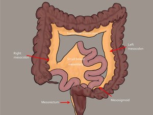
 Yellow line: right lateral peritoneal fold; 2) Red line: Toldt’s fascia (also known as anterior pararenal fascia) - Told's fascia is difficult to visualize, unless thickened within a pathological context. It is more easily visualized in the region where it was previously known as the anterior pararenal fascia, but it must be remembered that it occurs in every region of contact between peritoneum and retroperitoneum (even if it is not seen on imaging); 3) Purple region: small bowel mesentery; 4) Red region: transverse mesocolon; 5) Pink region: greater omentum - The transverse mesocolon and colon overlie the small bowel mesentery, and the greater omentum overlies the upper surface of the transverse mesocolon. Extensive adhesions occur between the under surface of the greater omentum and the upper surface of the transverse mesocolon; 6) Yellow region: right mesocolon; 7) Blue region: left mesocolon; 8) Orange region: retroperitoneum.")
Fig. 10:
Overview: 1) Yellow line: right lateral peritoneal fold; 2) Red line: Toldt’s...