ECR 2019 / C-0408
Percutaneous image-guided palliative cryoablation of bone metastases
Congress:
ECR 2019
Poster Number:
C-0408
Type:
Scientific Exhibit
Keywords:
Oncology, Interventional non-vascular, Musculoskeletal bone, Percutaneous, CT, Ablation procedures
Authors:
I. A. Burovik, G. Prokhorov, A. V. Mishchenko; Saint Petersburg/RU
DOI:
10.26044/ecr2019/C-0408

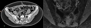
Fig. 3:
Metastasis in sacrum of ovarian cancer

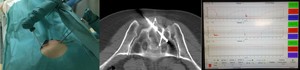
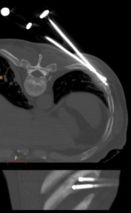
Fig. 4:
Procedure of cryoablation. Two cryoprobes and one needle microthermocouple....
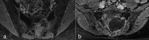
 and in 6 months after cryoablation (b). On postoperative study (b) cystic cavity in the zone of the lesion with slight enhancement of the walls can be seen")
Fig. 5:
MRI scans before (a) and in 6 months after cryoablation (b). On postoperative...

Fig. 6:
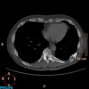
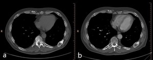
Metastasis in the rib of lung cancer

Fig. 7:
Probe positioning while cryoablation
 and in 8 months after cryoablation (b)")
Fig. 8:
Matastasis in the rib of lung cancer: before treatment (a) and in 8 months...