ECR 2019 / C-0943
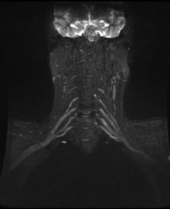
MRI of the brachial plexus: improved imaging technique.
Congress:
ECR 2019
Poster Number:
C-0943
Type:
Educational Exhibit
Keywords:
Inflammation, Hernia, Education and training, Technical aspects, Imaging sequences, Computer Applications-3D, Neural networks, MR-Diffusion/Perfusion, MR, Radiographers, Neuroradiology peripheral nerve, Anatomy
Authors:
A. P. Dominguez Castilla1, D. Sancho2, A. Merina2, C. Garcia Ansola2, J. BACHILLER EGEA2, L. LOPEZ RUIZ3; 1Villaviciosa de odon, M/ES, 2MADRID/ES, 3MADRID, casado/ES
DOI:
10.26044/ecr2019/C-0943

Fig. 7

Fig. 8

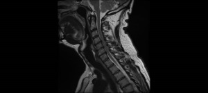
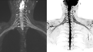
Fig. 3

Fig. 4

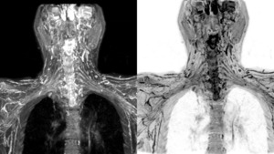
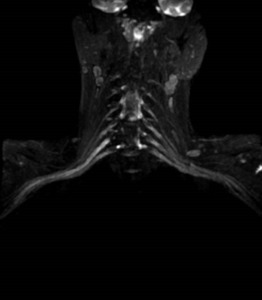
Fig. 9

Fig. 10

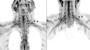
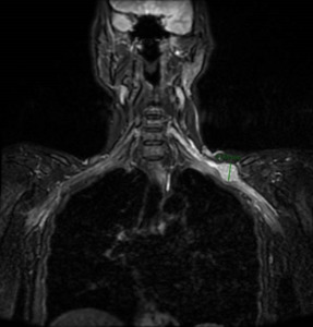
Fig. 11

Fig. 12

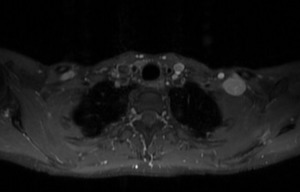
Fig. 13

Fig. 14