ECR 2019 / C-1017
Radiological evaluation of the lumbar lordosis in spine operated patients: is plain film still mandatory?
Congress:
ECR 2019
Poster Number:
C-1017
Type:
Scientific Exhibit
Keywords:
Outcomes, Comparative studies, MR, CT, Conventional radiography, Musculoskeletal system, Musculoskeletal spine
Authors:
A. Agustí, P. Garcia, I. García Duitama, G. salo bru, J. Ares, A. Solano; Barcelona/ES
DOI:
10.26044/ecr2019/C-1017

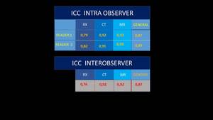
Fig. 6

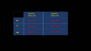
Fig. 7

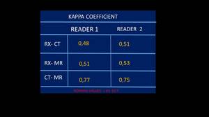
Fig. 8