A retrospective study of patients operated in our Hospital Center between 2012 and 2017 was carried out.
The impossibility of performing an adequate measurement of the lumbar lordosis in any of the radiological techniques used in the study was established as an absolute exclusion criterion,
regardless of the cause that generated it.
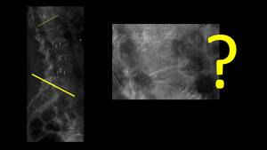
Fig. 2
The criteria for inclusion in the study were:
To be operated on an instrumented arthrodesis of the lumbar spine regardless of the number of levels and the cause of the intervention
Have a post-surgical radiological control by radiography and CT or MRI.
Arbitrarily,
it was defined that no more than 1 month should elapse between the radiograph and the other imaging tests.
The patient had to comply with the requirement to be operated in our Hospital and that the imaging tests were also performed in our center.
The study recruited a total of 240 patients (the first 40 patients who met the inclusion criteria each year).
The mean age was 64.9 ( max 94 min 19 ) years and the distribution was 56.6 percent of men and 43.4 of women.
83 patients had the three imaging techniques.
The measurement of lordosis in the lumbar curvature was performed according to the criteria defined by Robert Cobb in 1948,
establishing the angle between the sacral endplate and the cranial endplate of the vertebral transitional located between the lumbar curve and the thoracic curve.
It was defined that the transitional vertebra was always L1 in order to avoid dispersion in the values obtainedThe values of normality were established between the ages of 43 and 63.
The measurements made in the CT were obtained in the oriented MPR reconstructions.The measurements made in the MRI were obtained from T1WI or T2W2 images in the sagittal plane
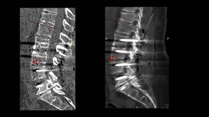
Fig. 3
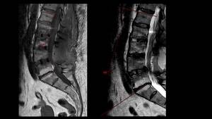
Fig. 4
All measurements were made in the PACS-IW centricity system as shown in the following image
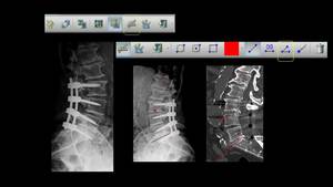
Fig. 5
Two experienced members of our unit performed the measurement of the Cobb angle according to the technique described above. Previously,
the two readers jointly measured three series of 10 patients.
Two measurements were made for each of the imaging techniques.The measurements in each of the techniques should not exceed 3 weeks
The person did not know the previous value of the angle obtained or the value of the angle obtained by the other reader. The numerical value of the measurement was obtained and established if it was within the range of normality or not.
Statistical analysis
To determine if any difference between measurement means was statistically significant,
calculated differences between means were statistically compared against the hypothetical value of zero.
The means,
standard deviations,
interclass and intraclass correlation coefficient and Kappa coefficient between the two readers and between the three measurements of each reader were calculated.The statistical management of the data included the coefficient of kappa with a p- value of 5%.


